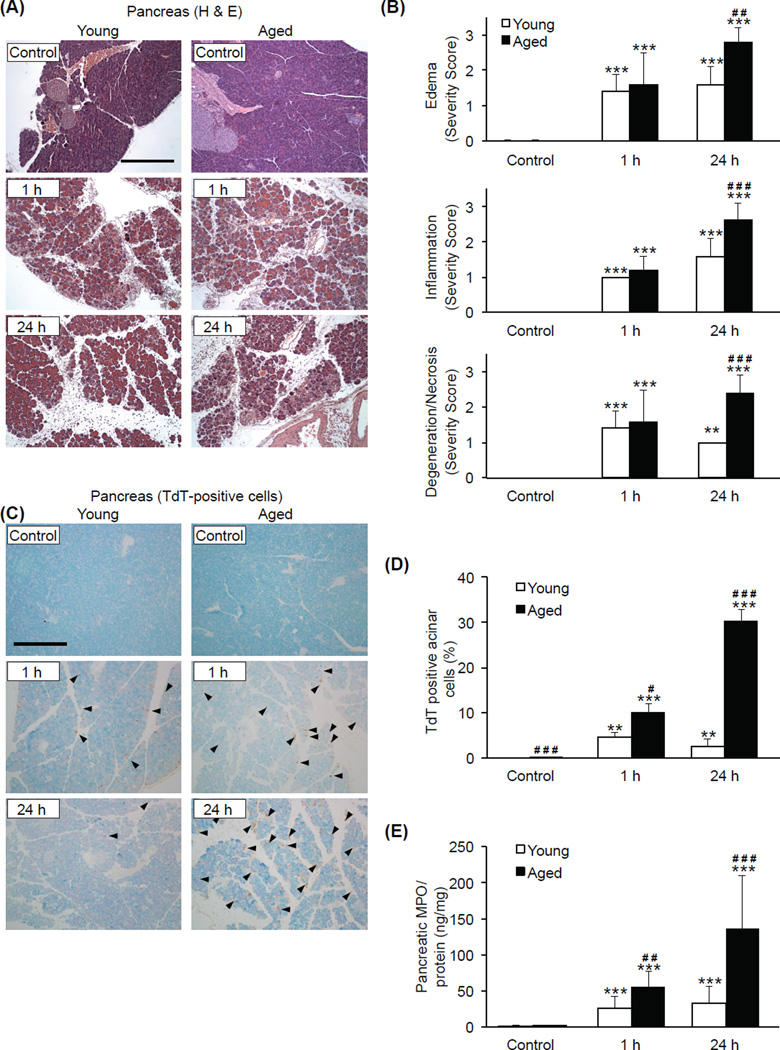
Figure 2. Age-associated increase in pancreatic tissue damage during AP.
Pancreatic tissue sections were obtained from young and aged mice sacrificed 1 or 24 h after the final (9th) caerulein injection. (A) Representative H&E-stained sections (original magnification, 100x, scale bar indicates 200µm). (B) Results of histological scoring. (C) Apoptotic cells indicated by immunohistochemical detection of TdT. Arrowheads represent TdT-positive cells (original magnification, 100x, scale bar indicates 200µm). (D) Acinar cell death expressed as a percentage of total acinar cell mass. (E) Pancreatic MPO levels. *** p<0.001, ** p<0.01 vs. control of the same age group; ### p<0.001, ## p<0.01, # p<0.05 vs. young mice at the same time point. n=5 in each group.

