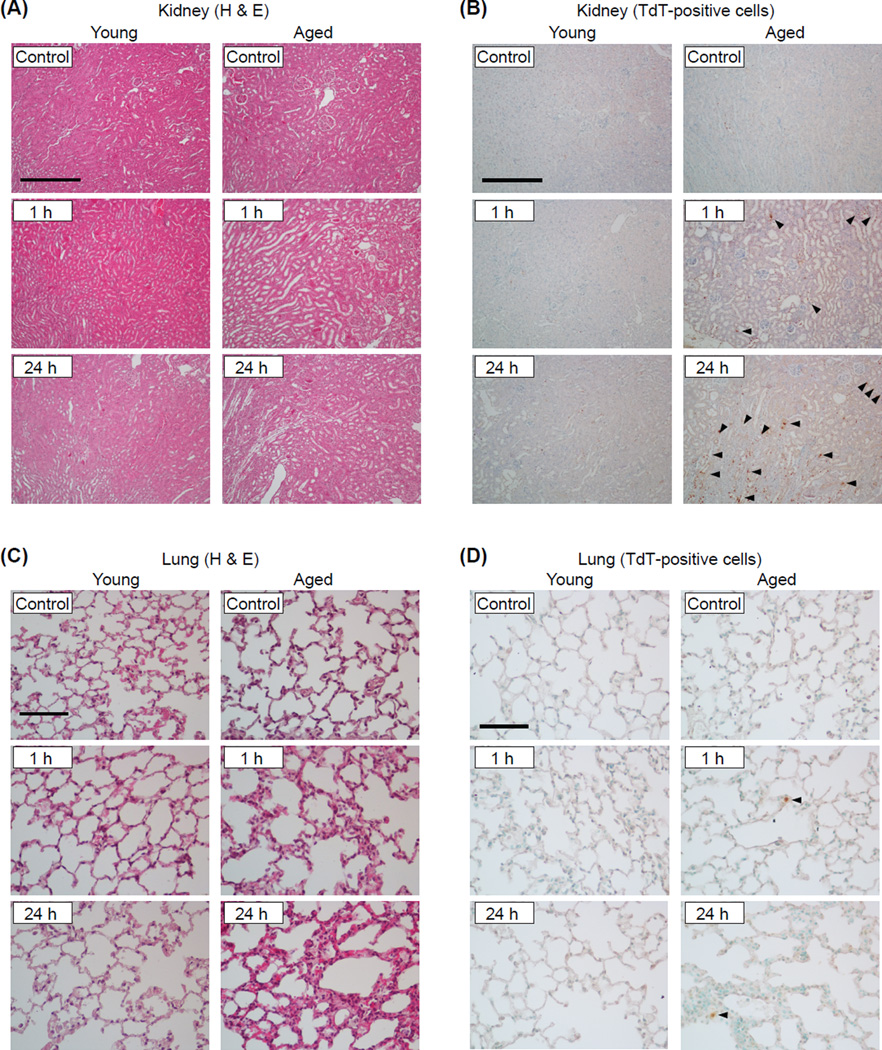
Figure 3. Age-associated severe tissue damage in the kidney and lung during AP.
Kidney and lung tissue sections were obtained from young and aged mice sacrificed 1 or 24 h after the final (9th) caerulein injection. The sections were either stained with H&E or processed for immunohistochemical detection of TdT-positive apoptotic cells. Representative H&E-stained kidney sections (A) and TdT-positive cells (indicated by arrowheads) in the kidney (B) (original magnification, 100x, scale bar indicates 200µm). Representative H&E-stained lung sections (C) and TdT-positive (indicated by arrowheads) in the lungs (D) (original magnification, 400x, scale bar indicates 50µm). n=5 in each group.

