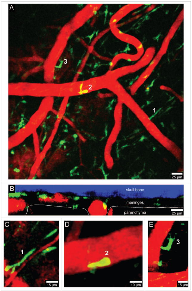
Figure 2.

Visualization of peripherally-derived myelomonocytic cells in LysM-GFP mice. A 3D time-lapse was captured through a thinned skull window of a naïve LysM-GFP mouse in a manner similar to that described in Figure 1. Monocytes, macrophages, and neutrophils (but not microglia) are visible in this transgenic mouse strain. Panel A (xy maximal projection) shows the distribution of myelomonocytic cells (green) in relation to blood vessels (red). Note in B (xz projection) that the LysM-GFP+ cells reside exclusively in the meningeal and perivascular spaces. Three cellular morphologies are depicted in panels C–E (labeled 1–3). Worm-like meningeal macrophages (C) are visible along meningeal blood vessels similar to those seen in CX3CR1-GFP+/- mice. On occasion, neutrophils / monocytes (D) can be observed patrolling blood vessels. In addition, amoeboid cells (E) are also visible around blood vessels (possibly perivascular macrophages). Skull bone is shown in blue. See Video S2.
