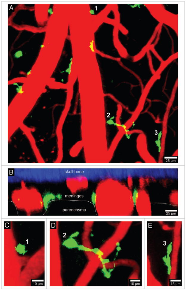
Figure 3.

Visualization of CD11c-YFP+ APCs. A 3D time-lapse was captured through a thinned skull window of naïve CD11c-YFP mouse in a manner similar to that described in Figure 1. The majority of CD11c-YFP+ cells (green) visible through a thinned skull reside in the meninges and perivascular spaces. Panels A (xy projection) and B (xz projection) show that CD11c-YFP+ cells are sparsely distributed in the meninges and perivascular spaces of a naïve mouse. There are three distinct cellular morphologies depicted in panels C–E (labeled 1–3). Small spheroid cells (C) that resemble monocytes are visible around blood vessels. There are also long stringy cells 50–75 μm in length (D) that are not completely juxtaposed to the vasculature but are instead intertwined with vessels. Lastly, juxtavascular CD11c-YFP+ cells (E) are visible that share some similarities (e.g., amoeboid) to the perivasular macrophages seen in LysM-GFP mice. Skull bone is shown in blue. See Video S3.
