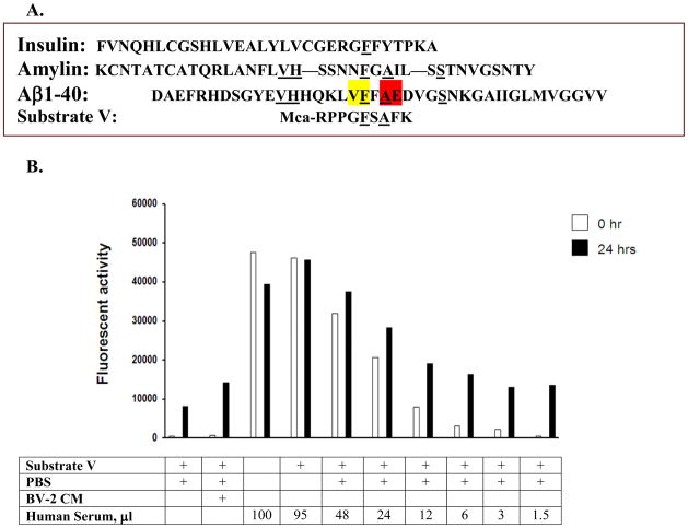
Figure 1. Serum activity of substrate V degradation.
The sequences of insulin, amylin and Aβ1–40 along with the sequence of substrate V are shown and compared (A). Cleavage sites of Aβ1–40 by IDE (yellow) and ACE (red) are illustrated. Different amounts of serum were incubated with substrate V at 37°C for time 0 and 24 hours (24 hr) and the degradation activity assessed by the fluorescence generation (B). BV-2 condition medium (CM) was used as a positive control.

