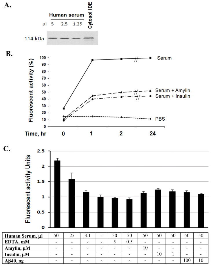
Figure 3. Characterization of IDE in human serum.
Decreasing amounts of serum were detected for IDE using the human IDE specific antibody, 6H9, in Western Blot, and the cytosol IDE was used as a positive control (A). 6 μl human serum was pre-incubated with or insulin (10 μM) or amylin (10 μM) at 37°C for 3 hours then followed by adding substrate V for continuation of different time points and the generated fluorescence were measured (B). IDE molecules from different amounts serum were immunocaptured by IDE antibody, 6A1, followed by adding and incubating with substrate V at 37°C for 3 hours, and the generated fluorescence was measured in the presence of different inhibitors. The background fluorescence without serum was used as the standard 1 and the times of enhanced fluorescence generated by the presence of serum relative to the background were calculated and illustrated. Insulin, amylin, Aβ1–40 or EDTA was applied to the assay to observe the specific inhibition of IDE (C).

