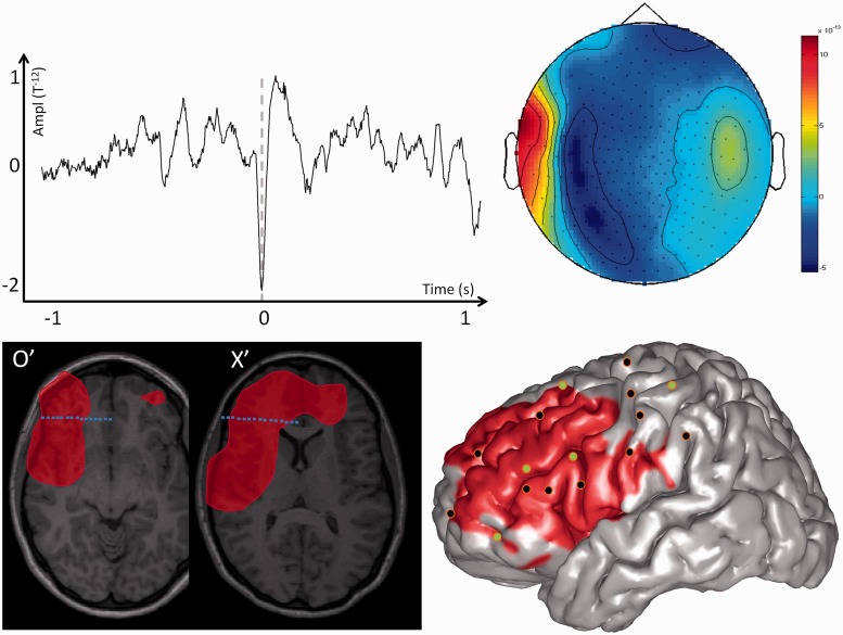
Figure 3.
MEG modelling of epileptic spikes and SEEG localization of the seizure-onset zone in a patient with a non-lateralized spiking volume. Top left: Example of one prototypical spike recorded with MEG for one single channel of Patient 7. Top right: Topographic map of the magnetic field recorded for all MEG channels at the main peak of the prototypical spike. Bottom left: The MEG spiking volume determined with VIES (red volume) is shown on representative MRI slices. On the same slices, the SEEG electrodes showing clear signal changes at seizure-onset are shown in blue, and electrodes without clear ictal activity are in white. Bottom right: SEEG implantation scheme of Patient 7. The location of the penetrating points of intracranial electrodes is determined by co-registering the patient’s post-implantation MRI onto the cortical mesh extracted from the MRI. The MEG spiking volume projected onto the cortical surface is shown as a red area. SEEG electrodes showing clear signal changes at seizure-onset are shown in green and electrodes without ictal changes are shown in black. Notice that the spiking volume is spatially very extensive and partly co-extensive with the seizure-onset zone determined with SEEG. The seizure-onset zone was considered as not perfectly delineated. However, because the epilepsy was very severe, the patient underwent surgical resection of left antero-mesial frontal cortex [frontal pole + anterior superior frontal gyrus (F1) + supplementary motor area and anterior cingulate gyrus] with an unsatisfactory surgical outcome (Engel III).

