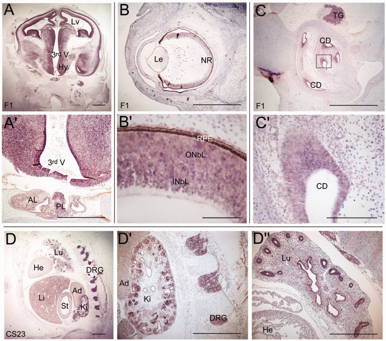
Figure 3.
Expression analysis of ARNT2 during human embryonic development. In situ hybridization using riboprobes against ARNT2 reveals expression in the developing brain and pituitary gland at 8 weeks of development (A). Strong expression can be seen in the hypothalamus and in the posterior lobe of the pituitary and weak expression is detected in the anterior lobe of the pituitary (A’). (B) Expression in the eye is detected in the developing neural retina (B’). (C) ARNT2 expression is detected in the inner ear, specifically in the cochlear ducts (C’). Expression is also observed in the trigeminal ganglion (C). (D) ARNT2 is expressed in the dorsal root ganglia, the developing kidney (D’) and in the lung, with strong expression in the bronchioles (D’’). Expression is also detected in the inner lining of the stomach. Scale bars: A, A’, B, D, D’ and D’’ = 100 µm; B’ = 10 µm; C = 50 µm; C’ = 20 µm. Lv = lateral ventricle; 3rd V = third ventricle; Hy = hypothalamus; PL = posterior lobe; AL = anterior lobe; Le = lens; NR = neural retina; INbL = inner neuroblastic layer; ONbL = outer neuroblastic layer; RPE = retinal pigmented epithelium; CD = cochlear duct; TG = trigeminal ganglion; Lu = lungs; He = heart; Li = liver; St = stomach; Ki = kidney; Ad = adrenal; DRG = dorsal root ganglia.

