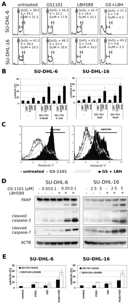
Figure 3. Induction of apoptosis in SU-DHL-6/16 cells after exposure to GS-1101 and LBH589.
(A) Cells were incubated with GS-1101 and/or LBH589 (LBH) for 48 h, and the cell cycle was analysed by flow cytometry. (B) sub-G0/G1 DNA content analysis of SU-DHL-6/16 cell lines after 48 h treatment. (C) Histogram of annexin V/7-aminoactinomycin D (7-AAD) positive cells, where annexin V/7-AAD positivity reflects the number of apoptotic (apo) and necrotic (nec) cells. Annexin V/7-AAD staining was employed to confirm the extent of apoptosis after the combined treatment. (D) Combination of GS-1101 and LBH589 significantly induce PAPR cleavage and activation of caspase-3, and -7 in SU-DHL-6/16 cells. Cells were treated for 24 h and protein extracts were analysed by Western Blot. (E) sub-G0/G1 DNA content analysis of SU-DHL-6/16 cells after 48 h combined treatment where SAHA substitutes LBH589 and GS-1101 was replaced by BKM120.

