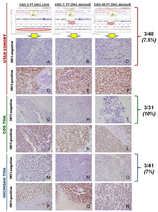
Figure 1. SMARCB1 (INI1/SNF5) LOH, Copy Number, and Immunohistochemistry (IHC) in Ewing Sarcoma.
The top three figures display the probe values and B-allele frequency for OncoScan™ FFPE Express assay, illustrating copy neutral LOH (A:EWS-5) and SMARCB1 homozygous deletions (B:EWS-7, C:EWS-40). IHC staining in the Utah cohort, COG tissue microarray (TMA), and Michigan TMA revealed SMARCB1 protein expression loss in 7–10% of Ewing sarcoma primary tumor samples. Utah cohort: A) Sample A contained SMARCB1 copy neutral LOH and shows loss of SMARCB1 (INI-1/BAF-47 antibody) nuclear expression in tumor cells. B–C) Samples B and C both had SMARCB1 homozygous deletions with loss of SMARCB1 nuclear expression in tumor cells. Note that samples A and C show retained SMARCB1 nuclear positivity in endothelial cells, which serves as an internal, positive control. D–F) All contained normal SMARCB1 copy number and LOH, and each sample shows diffuse SMARCB1 nuclear staining. COG TMA: G–I) Samples show loss of SMARCB1 nuclear expression in tumor cells. Note the retained SMARCB1 nuclear positivity in endothelial cells (Figure G, left). J–L) Samples show diffuse SMARCB1 nuclear staining. MICHIGAN TMA: M–O) Samples show loss of SMARCB1 nuclear expression in tumor cells. P–R) Samples show diffuse SMARCB1 nuclear staining.

