
Figure 3. RSK induces the expression of a coordinate motility and invasion gene program in MDCK cells.
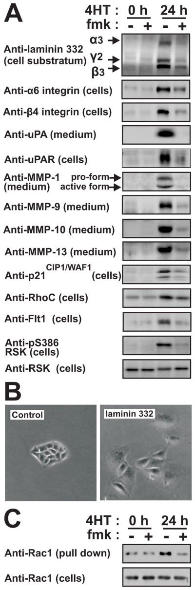
(A) Polarized MDCK-RAF1:ER cell monolayers were exposed to 1 μM 4HT and 6 μM fmk as indicated. After 24 h, the cells or the medium were analyzed by immunoblotting.
(B) Subconfluent MDCK cells were cultured in the absence or presence of 25 μM laminin 332 and photographed 48 h later.
(C) Polarized MDCK-RAF1:ER cell monolayers treated as in (A) were analyzed for active Rac1. All experiments were conducted 3–5 times with similar results.