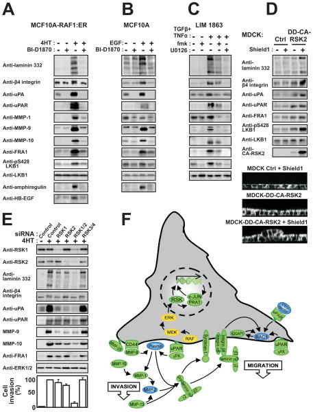
Figure 6. RSK stimulates a pro-motile/invasive gene program in various non-transformed or cancerous epithelial cell types.
MCF10A-RAF1:ER (A), MCF10A (B) and LIM 1863 cells (C) were exposed to 1 μM 4HT, 20 nM EGF, 10 ng/ml TNFα, 2 ng/ml TGFβ, 10 μM BI-D1870, 6 μM fmk or 10 μM U0126, as indicated. After 24 h, the cells or medium were analyzed by immunoblotting.
(D) MDCK cells expressing conditionally active CA-RSK2 or vector Ctrl were exposed or not to 1 μM of the inducer Shield1. The cells were analysed after 24 h by immunoblotting or assessed for multilayering after 72 h, as described in legend to Fig. 2A.
(E) MCF10A-RAF1:ER cells were subjected to siRNA knockdown of RSK1-4 in the combinations indicated. After 48 h, the cells were analysed by immunoblotting or in invasion assays, as described in the legend to Fig. 2D.
(F) The model summarizes the results of the present study and illustrates that RSK may induce mesenchymal invasive capacities in epithelial cells by stimulating a coordinate gene program (green) via FRA1-dependent and -independent mechanisms.

