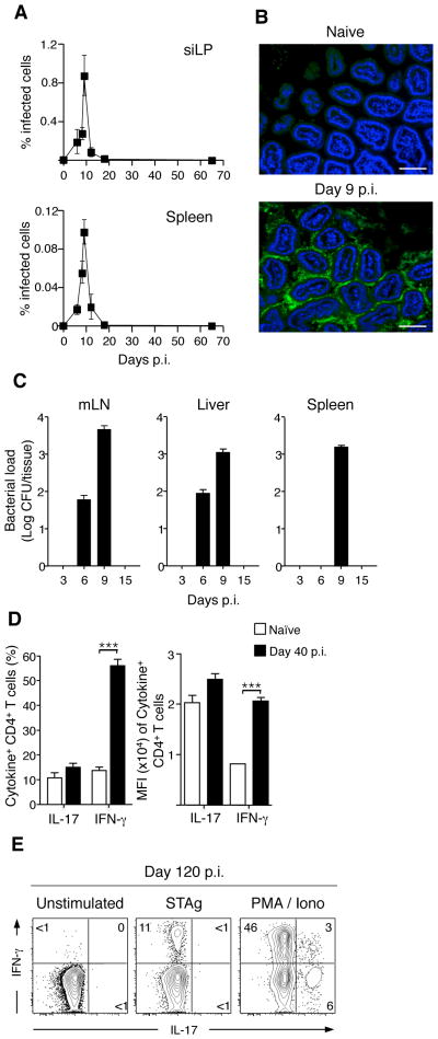
Figure 1. T. gondii infection leads to transient bacterial translocation but persistent lamina propria Th1 T cell responses.
(A) C57Bl/6 mice were infected orally with 15 cysts of RFP-expressing T. gondii. At the times indicated p.i., siLP and spleen were isolated and the percent of cells infected measured via flow cytometric analysis of RFP+ cells; N=1–8, n=3–15 mice per timepoint. (B) Small intestinal samples were isolated from naïve and day 9 T. gondii infected mice. Intestinal samples were then fixed, sectioned, counter-stained with DAPI and hybridized for FISH with a fluorescent probe specific to eubacterial 16S chromosomal sequences. Scale bar indicates 25 μM; N=7 (C) Counts of bacterial colonies cultured from spleen, mesenteric LN (mesLN) and liver at time points indicated post T. gondii infection; N=3, n=3–4 mice per timepoint. (D) Lymphocytes were prepared from the siLP of naïve or day 40 infected mice, stimulated with PMA/ionomycin and stained intracellularly for IFN-γ and IL-17. Bar graph shows the percent (Left) or the Mean Fluorescence Intensity (Right) of CD4+ cells expressing IFN-γ or IL-17. Shown is a representative example of three separate experiments; n=4–6 mice per experiment. (E) CD4 T cells were purified from the siLP by flow cytometry and co-cultured with bone-marrow derived DCs pre-loaded with Soluble T. gondii Antigen (STAg) or activated with PMA/ionomycin for 3.5 hours. Stimulated cells were stained intracellularly for IFN-γ and IL-17 and analyzed by flow cytometry. Shown is one of two experiments. Flow cytometry of CD4 cells is gated Live TCRβ+ CD4+ Foxp3−. Graphs show mean +/− SEM. ***p<0.001.

