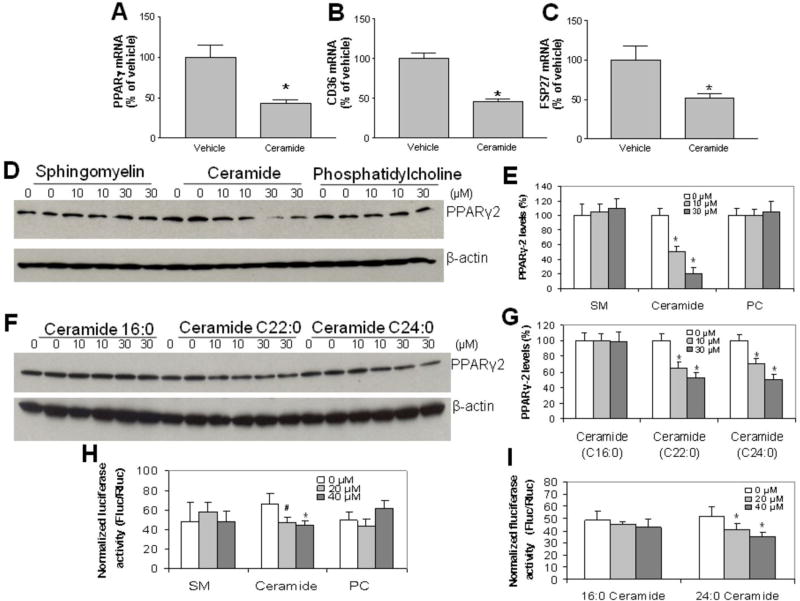
Figure 3. The exogenous supplementation of ceramide in culture significantly reducesPPARγ2 protein levels.
Huh7 cells were incubated with exogenous 0, 10, 30 μM of sphingomyelin, ceramide or phosphatidylcholine for 24 h. Cellular PPARγ2, CD36 and FSP27 mRNA levels were measured by real-time PCR. Panel A, PPARγ2 mRNA levels. Panel B, CD36 mRNA levels. Panel C, FSP27 mRNA levels. Cellular PPARγ2 mass were measured by Western blotting. Panel D and E, PPARγ2 Western blot after treatment with exogenous 0, 10, 30 μM sphingomyelin, ceramide or phosphatidylcholine for 24 h. Panel F and G, PPARγ2 Western blot after treatment with exogenous 0, 10, 30 μM of ceramides C16:0, C22:0, and C24:0 for 24 h. Panel H and I, PPAR reporter assay. Huh7 cells were transfected with PPAR reporter, negative control or positive control, respectively. After 24 h of transfection, the cells were treated with exogenous sphingomyelin, ceramide or phosphatidylcholine for 24 h, and then dual-luciferase reporter assay was performed. Panel H, dual-luciferase reporter assay after treatment with exogenous 0, 20, 40 μM of sphingomyelin, ceramide or phosphatidylcholine for 24 h. Panel I, dual-luciferase reporter assay after treatment with exogenous 0, 20, 40 μM of ceramides C16:0 and C24:0 for 24 h. Values are Mean ± SD., n=5, *P<0.05, **P<0.01.

