
Figure 2.
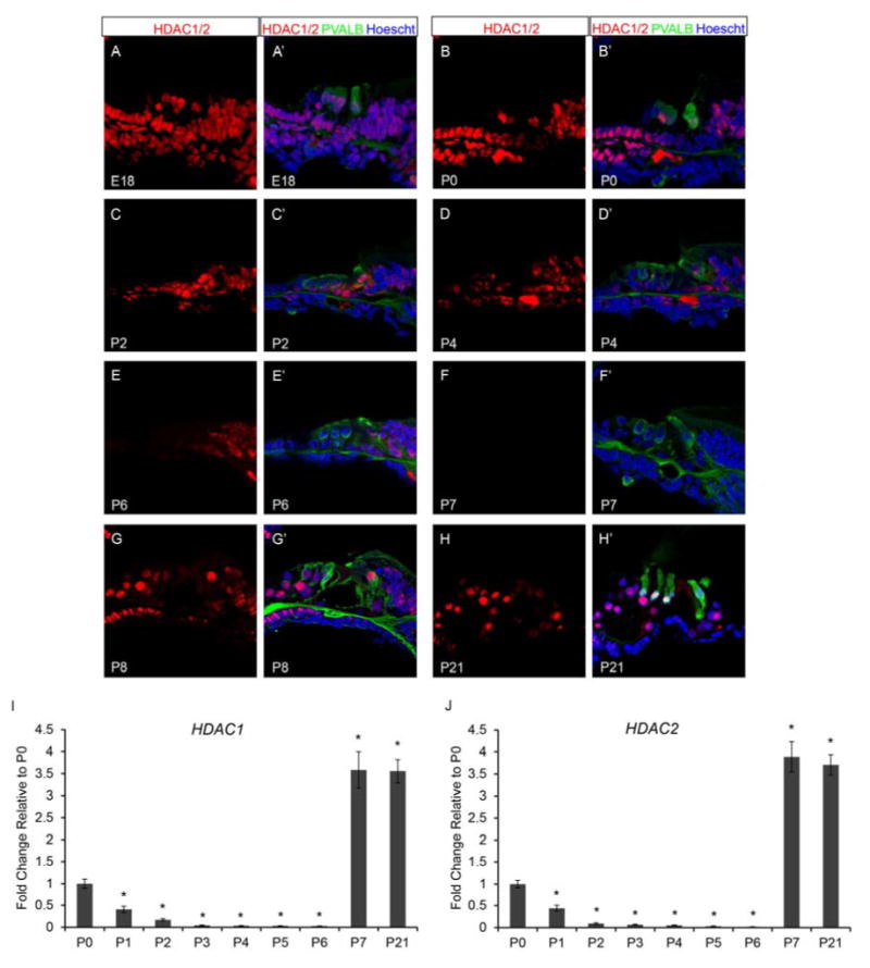
Neonatal murine organ of Corti expresses histone deacetylases, HDAC1 and HDAC2. Immunofluorescence of wild type neonatal mouse cochlea was performed using antibodies against HDAC1 and 2 (red), Parvalbumin (PVALB) (green), and Hoechst (blue). Taqman gene expression assays were done on microdissected organ of Corti. Expression levels are relative to endogenous gene controls 18S, GAPDH, and Actb. Fold changes are shown relative to P0. (A & A’) HDAC1/2 are found throughout the organ of Corti at E18.5. (B & B’) HDAC1/2 are still present in a majority of cell types in the organ of Corti at P0. (C-D’) Staining for HDAC1/2 appear reduced at P2 and P4 compared to E18.5 and P0. (E & E’) P6 organ of Corti retains some HDAC1/2 label in the OPCs, IPCs, IPhs, and IHCs. (F & F’) HDAC1/2 staining is no longer apparent in the organ of Corti by P7. (G-H’) HDAC1/2 reappears in the organ of Corti by P8 and continues to be detected at P21 (I) Expression of Hdac1 and Hdac2 are very similar at each time point analyzed. Hdac1 and Hdac2 expression steadily decreases from P0 to P6, then at P7 and P21 expression is increased compared to the P0 time point. All representative images were taken from the middle turn of the cochlea. *P<0.05 by Data Assist Software.