FIGURE 5:
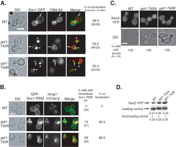
The YPT1-T40A mutation, like YPT1-T40K, confers Snc1-GFP accumulation in the ER but not endosome-to-Golgi transport defects. The three strains used here are YPT1 (WT), ypt1-T40K, and ypt1-T40A allele (as in Figure 1). (A) Half of the intracellular Snc1-GFP localizes to endosomes in ypt1-T40A and ypt1-T40K mutant cells. The endosomes of cells expressing Snc1-GFP were labeled with FM4-64, and cells were visualized using live-cell microscopy (as in Figure 3B). Left to right, DIC, GFP, FM4-64, merge, percentage colocalization of Snc1-GFP with endosomes (number of GFP puncta/number of cells visualized). Arrows point to GFP puncta that colocalize with FM4-64; arrowheads point to GFP puncta that do not colocalize with FM4-64. About 50% of the intracellular Snc1-GFP colocalizes with FM4-64 in ypt1-T40A and ypt1-T40K, as compared with ∼100% colocalization in wild-type cells. (B) Intracellular GFP-Snc1-PEM localizes to the ER in both ypt1-T40K and ypt1-T40A mutant cells. Cells expressing GFP-Snc1-PEM and the endogenously tagged ER marker Hmg1-mCherry (as in Figure 3A) were visualized by live-cell fluorescence microscopy. Left to right, DIC, GFP, mCherry, merge, percentage of cells with intracellular GFP-Snc1-PEM (N, number of cells visualized), and percentage colocalization (percentage of cells in which intracellular GFP-Snc1-PEM colocalizes with Hmg1). In wild-type cells all the GFP-Snc1-PEM is on the PM and does not colocalize with Hmg1 rings. In ypt1-T40A and Ypt1-T40K mutant cells, ∼90% of the intracellular GFP-Snc1-PEM colocalizes with Hmg1. (C) Kex2 recycling is not defective in ypt1-T40K and ypt1-T40A mutant cells. Endogenous Kex2-YFP was expressed in wild-type and ypt1-T40K and ypt1-T40A mutant cells. Top, cells were visualized by live-cell fluorescence microscopy (as in Figure 4C). Left to right, DIC, YFP, and percentage of cells with intracellular GFP puncta (N, number of cells visualized). (D) Immunoblot analysis of Kex2-YFP. Lysates from wild-type and ypt1-T40K and ypt1-T40A mutant cells expressing Kex2-YFP were subjected to immunoblot analysis using anti-GFP antibodies (as in Figure 4D). Bands were quantified and the ratio of Kex2 over the loading control is shown; ±, SD. Like wild-type cells, both ypt1-T40K and ypt1-T40A mutant cells show Kex2-YFP puncta and similar level of Kex2-YFP. Bar, 5 μm (A–C). Results represent at least two independent experiments.

