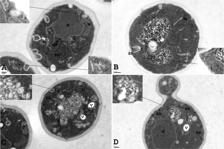
FIGURE 6:
Aberrant ER accumulation in ypt1-1 mutant cells overexpressing Snc1-GFP. Immuno-EM analysis was done with wild-type and ypt1-1 mutant cells expressing Snc1-GFP. Wild-type and ypt1-1 mutant cells expressing Snc1-GFP were grown to log phase, fixed, and processed for immuno-EM using anti-Hmg1 antibodies. Insets show enlarged view of ER structures labeled with gold particles. (A, B) In wild-type cells, Hmg1, an ER marker, is present exclusively on elongated ER membranes. (C, D) In ypt1-1 mutant cells Hmg1 is present on aberrant membrane structures. ER, endoplasmic reticulum; M, mitochondria; N, nucleus; V, vacuole. Bar, 200 nm. Results represent two independent experiments; quantification is shown in Supplemental Table S1.

