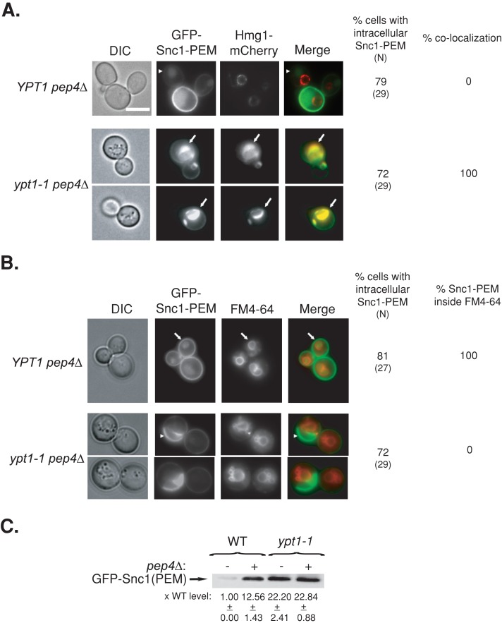
FIGURE 7:
GFP-Snc1-PEM accumulates in the vacuole of pep4∆, but not ypt1-1 pep4∆, mutant cells. (A) Accumulation of intracellular GFP-Snc1-PEM outside the ER of YPT1 (WT) pep4∆ cells defective in vacuolar degradation. GFP-Snc1-PEM was expressed from a 2 μ plasmid and mCherry-Hmg1 from its endogenous locus (as in Figure 4A) in YPT1 pep4∆ and ypt1‑1 pep4∆ mutant cells. Cells were visualized by live-cell fluorescence microscopy. Left to right, DIC, GFP, mCherry, merge, percentage of cells with intracellular GFP-Snc1-PEM (N, number of cells visualized), and percentage colocalization (percentage of cells in which intracellular GFP-Snc1-PEM colocalizes with Hmg1-mCherry). In YPT1 (WT) pep4∆ cells, intracellular GFP-Snc1-PEM does not colocalize with the Hmg1 rings (arrowheads). In ypt1-1 pep4∆ double mutant cells, intracellular GFP-Snc1-PEM colocalizes with Hmg1 (arrows). (B) Intracellular GFP-Snc1-PEM accumulates inside the vacuole of YPT1 pep4∆ but not ypt1-1 pep4∆ mutant cells. The vacuolar membrane of YPT1 pep4∆ and ypt1‑1 pep4∆ mutant cells expressing GFP-Snc1-PEM was labeled with FM4-64. Cells were visualized by live-cell microscopy. Left to right, DIC, GFP, FM4-64, merge, percentage of cells with intracellular GFP-Snc1-PEM (N, number of cells visualized), and percentage localization of intracellular GFP-Snc1-PEM inside the vacuole. Arrows point to GFP localized inside FM4-64-labled vacuoles YPT1 (WT) pep4∆ cells; arrowheads point to GFP that does not localize inside FM4-64-labled vacuoles in ypt1-1 pep4∆ double mutant cells. (C) Accumulation of GFP-Snc1-PEM in ypt1-1 mutant cells is not dependent on the vacuolar Pep4 protease. The protein level of GFP-Snc1-PEM in lysates of wild-type and ypt1-1 mutant cells with or without pep4∆ was determined using immunoblot analysis and anti-GFP antibodies. Bands were quantified, and the ratio of GFP-Snc1-PEM in mutant versus wild-type cells is shown at the bottom; ±, SD. The level of GFP-Snc1-PEM is higher in pep4∆ mutant cells than in wild-type cells, indicating that the protein is degraded in the vacuole. In contrast, the high level of GFP-Snc1-PEM In ypt1-1 mutant cells is not dependent on pep4∆, indicating that the protein does not reach the vacuole. Bar, 5 μm. Results represent at least two independent experiments.

