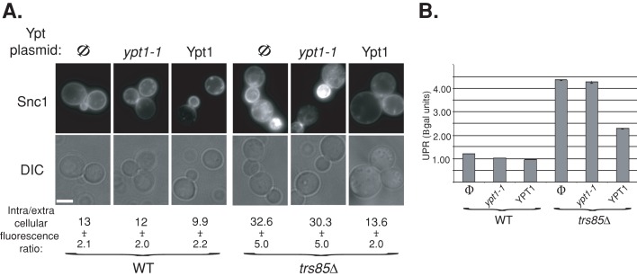
FIGURE 9:
A role for the Trs85-Ypt1 interaction in ER-phagy. (A) Overexpression of Ypt1 suppresses the Snc1-GFP accumulation defect of trs85∆ mutant cells. Top, wild-type (left) and trs85∆ mutant cells (right) expressing Snc1-GFP from a 2 μ plasmid were transformed with a second plasmid expressing Ypt1-T40K (mutant) or Ypt1 (wild-type) proteins (Φ, empty vector control). Cells were visualized by live-cell microscopy; representative cells are shown: GFP (top) and DIC (bottom). Bar, 5 μm. Bottom, quantification of intracellular/extracellular fluorescence ratio. The ratio of fluorescence inside and outside cells was determined using ImageJ (40 cells for each strain). The level of intracellular fluorescence in trs85∆ mutant cells is three times higher than in wild-type cells, and this increase is suppressed if Ypt1 but not Ypt1-T40K mutant protein is overexpressed. (B) Overexpression of Ypt1 suppresses UPR induction in trs85∆ mutant cells. Cells were transformed with three plasmids expressing LacZ under a UPR promoter, Snc1-GFP, and Ypt1 (Φ, Ypt1, or Ypt1-T40K). Induction of the UPR was determined in cell lysates using the βGAL assay (as in Figure 8B). UPR is induced in trs85∆ mutant cells (Φ) when compared with wild type, and this induction is partially suppressed by overexpression of Ypt1 but not Ypt1-T40K protein. Results represent at least two independent experiments; error bars, SD.

