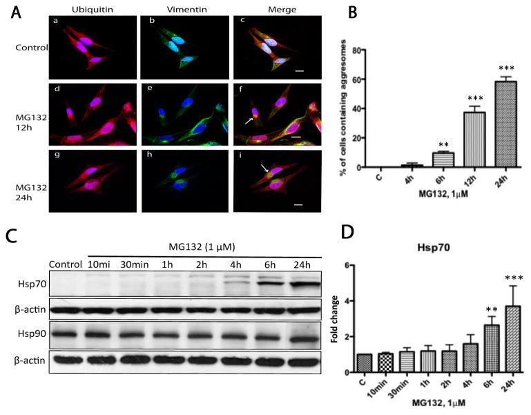
Figure 5. Prolonged treatment with MG132 resulted in aggresome formation.
(A) Aggresome formation was examined by confocal microscopy in N27 cell treated with MG132 (1μM) for 24h. Aggresomes were characterized as vimentin-caged, ubiquitin-positive perinuclear inclusions. For these studies cells were either treated with DMSO (a–c) or MG132 (1μM, d–f) for 2h to 24h, then processed for confocal laser microscopy using double immuno-labeling with antibodies to ubiquitin (red) and vimentin (green). Nuclei were stained with DAPI (blue). Arrows (panels f, i) indicate aggresome-like inclusion bodies. In these studies no aggresomes could be found at early time points (2 and 4h) after treatment with MG132. (B) Quantitative determination of aggresome formation. Cells were treated with MG132 (1μM) and at the indicated times the percentage of aggresome containing cells were determined by counting 100 cells across 20 random fields. The data represents the means ± SD, (n=3). Scale bar 20μm. (**p<0.01 and ***p<0.001, ANOVA using Dunnet’s post test). (C) Induction of Hsp70 but not Hsp90 following treatment with MG132. N27 cells were exposed to MG132 (1μM) from 10min to 24h as indicated and cytosolic fractions were then analyzed by immunoblot analysis for Hsp70 and Hsp90. (D) Fold changes of Hsp70 levels are normalized by control and estimated by densitometry. These data are presented as mean ± SD, (n=3); **p<0.01 and ***p<0.001 are considered significant by ANOVA using Dunnet’s multiple comparison test.

