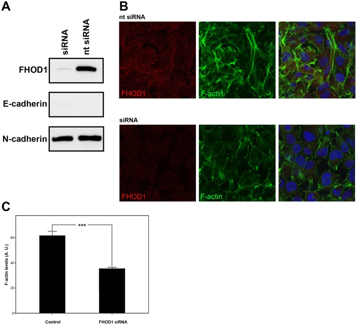
Figure 4. FHOD1 siRNA knockdown changes the morphology of UT-SCC-43B cells and reduces the number of stress fibres.
A) Western blotting shows that FHOD1 siRNA treatment significantly reduces FHOD1 expression. The cells are still E-cadherin negative and the expression of N-cadherin is unaltered. B) Cells treated with non-targeting siRNA (upper panel) express FHOD1 and have a mesenchymal phenotype. Stress fibres are abundant. FHOD1 siRNA abolishes FHOD1 staining (lower panel). siRNA treated cells have less actin stress fibres and are morphologically rounder and flatter. Nuclei are stained with DAPI (blue). C) F-actin staining is significantly reduced in FHOD1 siRNA treated cells. Phalloidin staining intensity is reduced by 49%. Bars indicate standard error of mean. *** p<0.0001. AU = arbitrary units.

