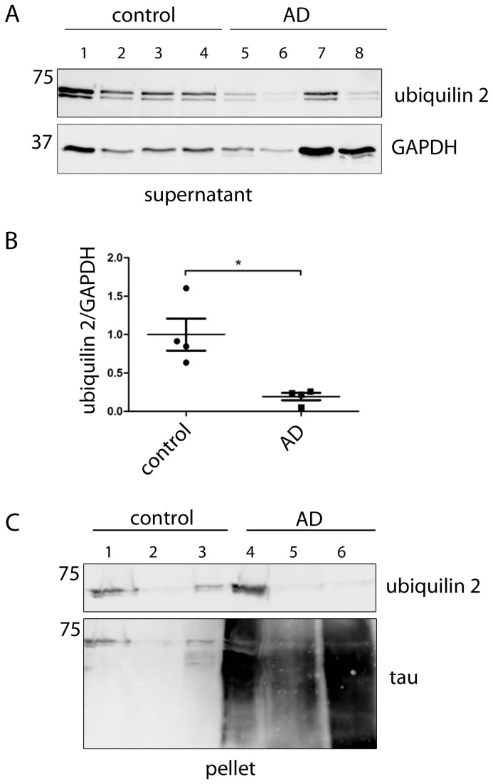
Figure 3. Ubiquilin 2 levels are decreased in AD patients.
(A) Western blot analysis of brain lysates from AD (Braak stage 5-6) and control (Braak stage 1) brain material using an ubiquilin 2 antibody and a GAPDH antibody as loading control. (B) Quantification of Western blot signals in (A). Ubiquilin 2 signals were normalized to GAPDH and statistically analysed (Mann–Whitney test, * p <0.05). (C) Supernatant and pellet lysate fractions of brain material from 3 AD patients (Braak stage 6) and 3 controls (Braak stage 1) were analysed by Western blotting. Pellets were treated with formic acid to dissolve aggregates prior to Western blotting. GAPDH was used as loading control (supernatant); tau was used as a control for successful extraction of the aggregates in the AD samples (pellet). AD, Alzheimer’s disease.

