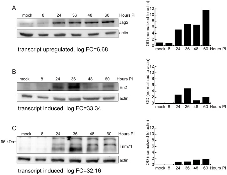
Figure 6. Validation of RNA-Seq analysis of host genes by western blot.
(A) Immunoblot analysis of MEF.K (A) or Balb/c MEF (B–C) cell lysates infected with wild-type MCMV. Cell lysates were separated by SDS-PAGE, transferred to PVDF membrane, and probed with antibody to Jag2 (A), EN2 (B) or Trim71 (C). Monoclonal antibody to actin was used as loading control. Bar charts represent relative quantification of proteins. In the case of Trim71 (C where anti-Trim71 antibody detected multiple bands, the bars show quantification of the middle band.

