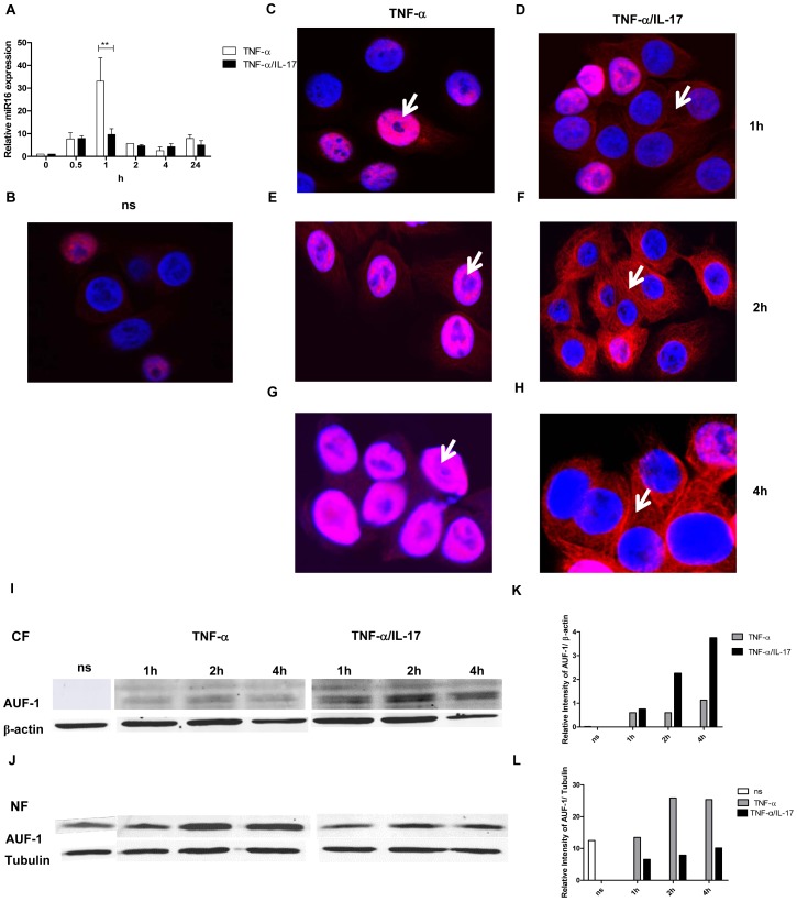
Figure 5. IL-17 limits miR16 expression and promotes cytoplasmic localization of AUF-1.
a) Q-PCR showing time dependent expression of miR16 measured in NCI-H292 cells stimulated with TNF-α or TNF-α plus IL-17. miR16 is presented relative to that of unstimulated, non-transfected cells. b–h) Representative confocal images showing the localization of AUF-1 in resting NCI-H292 cells (b) and in cells stimulated with TNF-α (c, e, g) or TNF-α plus IL-17 (d, f, h) for 1, 2 and 4 h respectively as described in methods. The nucleus is stained with DAPI. White arrows indicate the predominant localization of AUF-1. Two experiments were performed with similar results. i, j) Western Blot analysis of AUF-1 in the cytoplasmic (i) and nuclear (j) fractions of resting cells (ns) or cells stimulated with TNF-α or TNF-α plus IL-17. β-actin and tubulin are markers for the cytoplasmic and nuclear fractions respectively. k, l) Quantitative analysis of AUF-1 normalised to β-actin in cytoplasmic fraction (k) and to tubulin in nuclear fraction (l) obtained from the Western Blot.

