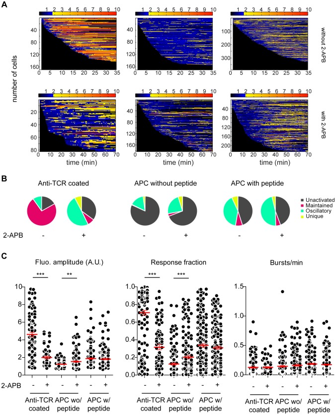
Figure 7. Naive CD4+ T cells display mainly intracellular calcium oscillations upon antigenic challenge. (A) Barcoding of stimulation.
The fluorescence intensity of all loaded and responsive murine 3A9 naive CD4+ T cell acquired by confocal microscopy and detected by MAAACS are normalized and displayed as a barcoded response over time (color-coded as indicated). Anti TCR antibody coated surface (left panel): Cells were seeded onto anti TCR antibody coated Lab-tek chambers with (ncells = 117) or without 2-APB (ncells = 276). Antigen presenting cell (middle and right panel): Cells were seeded onto COS-7 experimental antigen presenting cells loaded or not with HEL peptides (see materials and methods) in the presence of 2-APB (ncells = 266, without peptide; ncells = 112, with peptide) or without 2-APB (ncells = 491, without peptide; ncells = 349 with peptide). (B) Calcium response modes in naive CD4+ T cells represented in pie charts (unactivated in gray, maintained in pink, oscillating in green and unique in yellow). (C) Naive CD4+ T cell calcium responses upon different stimuli revealed by MAAACS. Left panel - fluorescence amplitude of T cells in the absence or presence of 2-APB. Middle panel - The response fraction of T cells in the absence or presence of 2-APB. Right panel - frequency of calcium bursts in T cell in the absence or presence of 2-APB. Scatter plots only show signals from responding cells (THact = 1.74 without 2-APB, THact = 1.89 with 2-APB). The mean value +/− SEM is represented in red. Statistical tests were carried out with the Mann Whitney non-parametric test (*** = p<0.001; ** = 0.001<p<0.01; * = 0.01<p<0.05; ns = p>0.).

