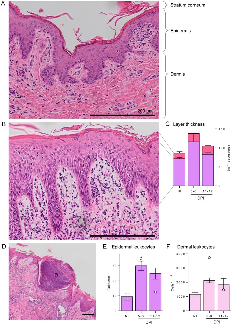
Figure 2. Leukocyte infiltration and thickness of buffalo skin following infection.
A buffalo skin section from non-infected buffaloes (A) are compared to those following moderate infection with S. japonicum cercariae (B and D), which resulted in a large influx of leukocytes in dermal and epidermal layers (B) and granulocytic abscesses (a) formation (D). Stratum corneum, epidermis and dermis are indicated for reference. Staining was performed with haematoxylin and eosin, and all scale bars represent 200 µm. Leukocytes were enumerated in the epidermis (E) and dermis (F) in non-infected (NI) and 5–6 or 11–12 days post-infection (DPI) buffaloes. For each buffalo 5 fields of view of the dermis at 400× magnification were enumerated and averaged, while for the epidermis 5 lengths of 0.26 mm were enumerated and averaged. The thicknesses of the epidermis (purple) and stratum corneum (pink) (C) were measured by determining volume of each layer relative to the length of the skin section, and 3 sections of 0.8 mm were averaged for each individual. Bars represent the mean of the moderate infections with standard error; the two high infections are shown in E and F as white dots. Significance above NI levels is shown by the asterisk (p = 0.015).

