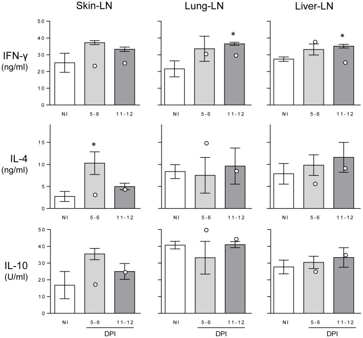
Figure 5. Cytokine levels in stimulated lymph node cell culture supernatants.
Levels of IFN-γ, IL-4 and IL-10 were measured by ELISA from PMA/ionomycin stimulated lymphocytes taken from skin-, lung- and liver-lymph nodes (LN) from non-infected (NI) and 5–6 or 11–12 days post-infection (DPI) buffaloes. Bars represent the mean of the moderate infections with standard error; the two high infections are shown as white dots). Asterisks indicate significance above NI levels (p<0.05).

