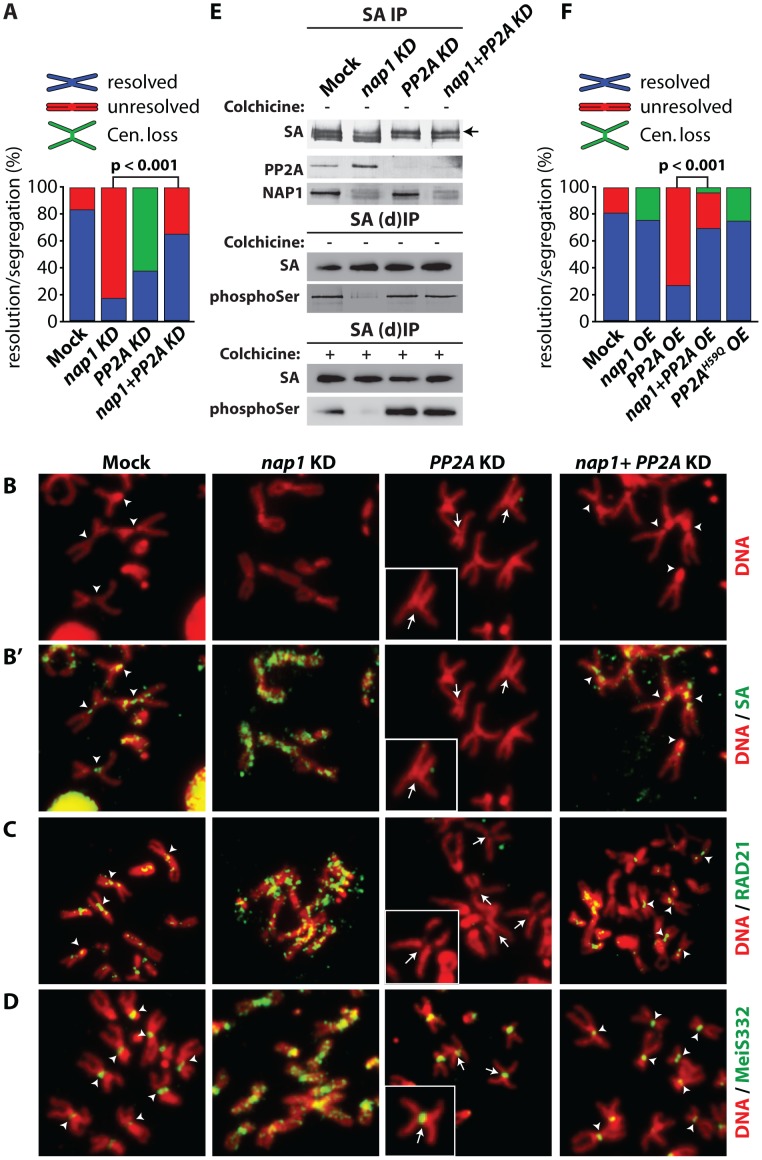
Figure 7. NAP1 and PP2A act antagonistically in cohesin cycle.
(A) Analysis of mitotic chromosomes from colchicine-treated S2 cells after knockdown of NAP1, PP2A or both factors. We quantified the frequency of resolved (blue), unresolved (red) sister chromatids and loss of centromeric cohesion (Cen. Loss; green). Concomitant depletion of NAP1 and PP2A resulted in a statistically significant increase of the frequency of resolved chromatids compared to the NAP1 knockdown, as determined by χ2-test (n>30, from 3 biological replicates). For the corresponding Western blot analysis see Figure S7A. (B) Representative example of mitotic chromosomes from colhicine-treated S2 cells depleted for NAP1, PP2A or for both proteins. DNA visualized by DAPI staining is shown in red. Centromers are indicated by arrowheads, whereas loss of centromeric cohesion is indicated by full arrows. (B′) The localization of SA (green) on mitotic chromosomes same as in (B) was determined by indirect immunofluorescence. (C) RAD21 (green) localization on mitotic chromosomes. (D) MeiS332 (green) localization on mitotic chromosomes. (E) Depletion of PP2A restores SA phosphorylation in cells lacking NAP1. Western blot analysis of SA IPed from either mock-treated S2 cells or after knockdown (KD) of NAP1, PP2A or both proteins under normal (top panel) or denaturing (middle panel, (d)IP) conditions from asynchronously dividing cells (− colhicine) or colhicine treated cells (bottom panel, + colhicine). Blots were probed with antibodies against SA, phosphorylated serine, PP2A or NAP1. After NAP1 knockdown, SA phosphorylation levels drop substantially. Whereas depletion of PP2A alone does not affect bulk SA phosphorylation, concomitant knockdown of PP2A and NAP1 neutralized the effect of NAP1 depletion, leading to restored levels of phosphorylated SA. Antibodies against phosphoSer recognize a band corresponding to the migration of SA. A slower migrating form of SA, presumably due to phosphorylation, is indicated by an arrow. (F) Analysis of mitotic chromosomes from colchicine-treated S2 cells after over-expression (OE) of GFP (Mock), NAP1, PP2A, both NAP1 and PP2A or the catalytic mutant PP2AH59Q. Quantification of mitotic phenotypes was as described above (A). Overexpression of PP2A, but not PP2AH59Q, resulted in significant increase of the frequency of unresolved chromatids. The PP2A over-expression phenotype was rescued by co-expression of NAP1, as determined by χ2-test (n>30, from 3 biological replicates). For the corresponding Western blot analysis see . Representative examples of mitotic chromosomes are shown in Figure S7C–D.

