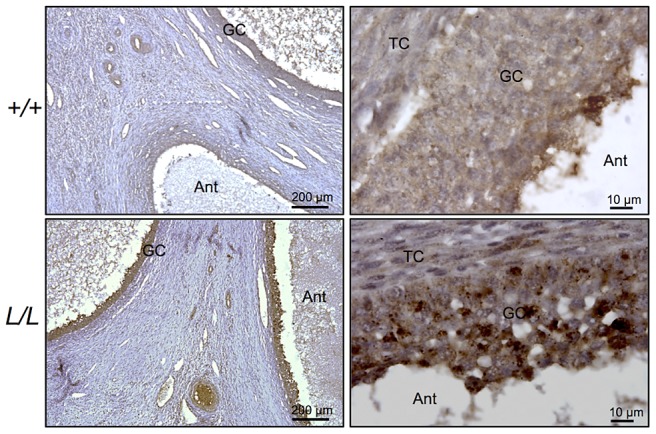
Figure 3. Immunostaining for B4GALNT2 in Lacaune sheep ovary.

Photomicrographs of ovarian sections from +/+ and L/L ewes stained with anti-B4GALNT2 rabbit polyclonal antibody (1/50 dilution). Sections were counterstained with hematoxylin. A black segment indicates the microscopy magnification scale. GC, granulosa cell layer; TC, theca cell layer; Ant, antral cavity.
