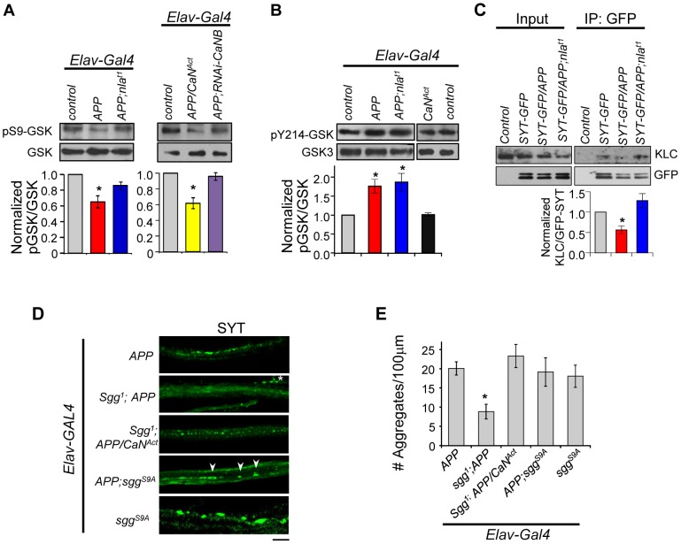
Figure 10. APP upregulation triggers changes in GSK-3β signaling downstream of calcineurin and affects synaptotagmin-kinesin interaction.
(A) Western blots performed using antibody specific for GSK-3β phosphorylated on Ser9 (pS9-GSK) and total GSK-3β. (B) Western blot using antibody specific for GSK phosphorylated at Tyr214 in Drosophila (pY214-GSK). For (A) and (B) quantification of the ratio of phosphorylated GSK and GSK-3β was normalized to control. n≥3 for each, * p<0.05. (C) Western blots showing reduced interaction between SYT-GFP and kinesin light chain (KLC). Control indicates parallel immunoprecipitation performed using flies that do not express SYT-GFP. n = 3, * p<0.05. (D) Images of 3rd instar larval motor axon stained with synaptotagmin. Arrowheads highlight aggregates and * indicates background staining coming from synaptotagmin in the NMJ. Scale bar: 10 µm. (E) Quantification of the number of aggregates found in each genotype. All values are mean ± S.E.M, * p≤0.05 compared to APP overexpression. n≥4 independent experiments per genotype.

