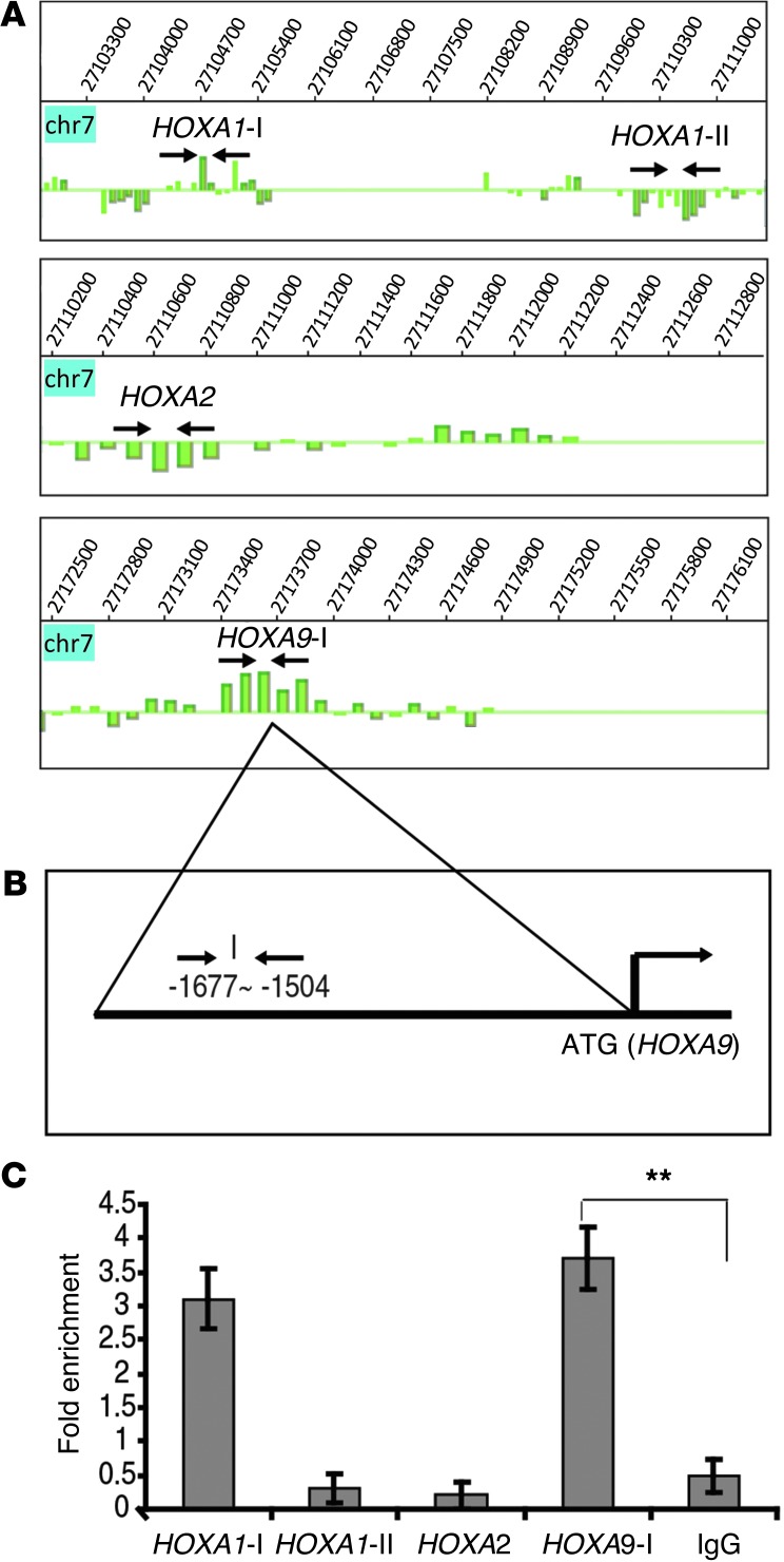
Figure 4. SALL4 binds to the promoter region of HOXA9 in the AML cell line.
(A) Analysis of ChIP-chip data showing SALL4 binding sites at the promoter regions of HOXA genes. Arrows indicate location of primers for validation by ChIP-qPCR. (B) Human HOXA9-I promoter region. (C) SALL4 bound to the promoter region of HOXA9 in KG1a cells, as evaluated by ChIP-qPCR. All values represent the average of at least 2 separate pulldowns (biological duplicates) and qPCR assays. Standard error bars were calculated from the SD of separate trials (biological repeats). Promoter regions from HOXA1-II and HOXA2 were used as internal negative controls, the region for HOXA1-I was used as a positive control, and rabbit IgG was used as a negative control for antibody specificity. **P < 0.001.

