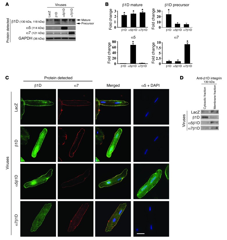
Figure 1. Combined expression of integrin α and β subunits is required for highest level of cell surface expression of integrins.
(A) Expression of integrins in ARVMs 48 hours after adenovirus infection. Western blot detecting LacZ, β1D integrin (β1D), α5 integrin (α5), and α7 integrin (α7) indicates increased expression of respective integrin subunits following viral infection. (B) Densitometric quantification of integrin expression in virally infected cells. Note that immature (precursor) form of β1D integrin subunit is increased when the cells are infected with the β1D subunit alone but not when they are infected with both α and β subunits. *P < 0.05 vs. LacZ, n = 6 per group (1-way ANOVA). (C) Representative immunofluorescent images of CMs following adenoviral-mediated OE of LacZ (control), β1D integrin alone, or β1D integrin in combination with α5 integrin or α7B integrin. β1D expression is noted on the cell membrane as well as in the cytoplasm when the β1D integrin subunit is overexpressed alone. When combined OE of α and β subunits occurs, the integrin expression is dominantly at the membrane, with little detected in the cytoplasm. Antibodies detect β1D (green), α7 (red), or α5 (green). Nuclei were stained with DAPI (blue). Scale bar: 20 μm. (D) Western blot analysis of β1D integrin expression in cytosolic and cell membrane fractions in ARVMs. As confirmation of the microscopic data, Western blotting shows that β1D integrin singly infected cells expressed integrin dominantly in the cytosolic fraction, while combined αβ integrin subunit infection allowed dominant cell membrane fraction expression.

