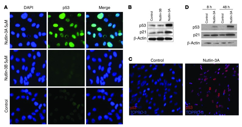
Figure 3. Nutlin-3 induces p53 expression and a downstream target in HUVECs and HUVSMCs.
(A) HUVECs were seeded on plastic-bottom culture dishes precoated with gelatin and treated with either 5 μM of Nutlin-3A, 5 μM of Nutlin-3B, or vehicle after 8 hours. Images were taken with an epifluorescent microscope and are representative of p53 expression (middle column) in the nucleus (left column) of HUVECs. Original magnification, ×400. (B) Western blot analysis for p53 and p21 was performed on lysates obtained from HUVECs treated with Nutlin-3A, Nutlin-3B, or vehicle in various concentrations for 8 hours. (C) Representative magnified confocal images are shown of HUVSMCs treated with either 7.5 μM of Nutlin-3A or vehicle after 8 hours. Original magnification, ×200. Nuclear p53 expression is shown in red and TO-PRO-3, a nuclear stain, in blue. (D) Western blot analysis for p53 and p21 was performed on lysates obtained from HUVSMCs treated with Nutlin-3A or vehicle.

