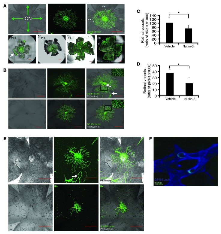
Figure 7. Nutlin-3 inhibits postnatal retinal vascular proliferation.
(A) Postnatal mouse retinal vascular development after birth (upper left panel). The optic nerve (ON) at the center of a retinal whole mount with green arrows indicating the direction of postnatal blood vessel growth (upper middle and right panels). GS-IB4 lectin staining of a retinal whole mount in the developing mouse pup demonstrating radial growth pattern of postnatal development of retinal vasculature. (lower panels). Double asterisks indicate areas of avascular retina. (B and C) Neonatal mice were given subcutaneous injections in the nape. (B) The retinal vasculature is abrogated in the Nutlin-3–treated eyes (n = 6) (bottom row) compared with the sham-injected mice (n = 4) (middle row). In addition, we noticed loss of smaller caliber vessels (inset, right column) in the Nutlin-3–treated mice. (C) The graph represents retinal vasculature measured as a function of pixels compared with the amount of retinal tissue in each mouse eye (*P < 0.05, representative of 2 independent experiments). (D and E) Neonatal mice were injected in the periocular area of each eye. (E) Nutlin-3–treated (lower row) mice (n = 8) have less retinal vasculature compared with sham-injected mice (n = 8) (upper row). (D) Same quantification method used in B shows a statistically significant difference between the 2 groups (*P < 0.01, representative of 2 independent experiments). (F) Rare TUNEL-positive cells identified in the retinal vasculature of a Nutlin-3–treated mouse. Original magnification, ×400. White arrows point to residual fetal vasculature unable to be removed during dissection. Data represent mean ± SD. *P < 0.05. Scale bars: 500 μm.

