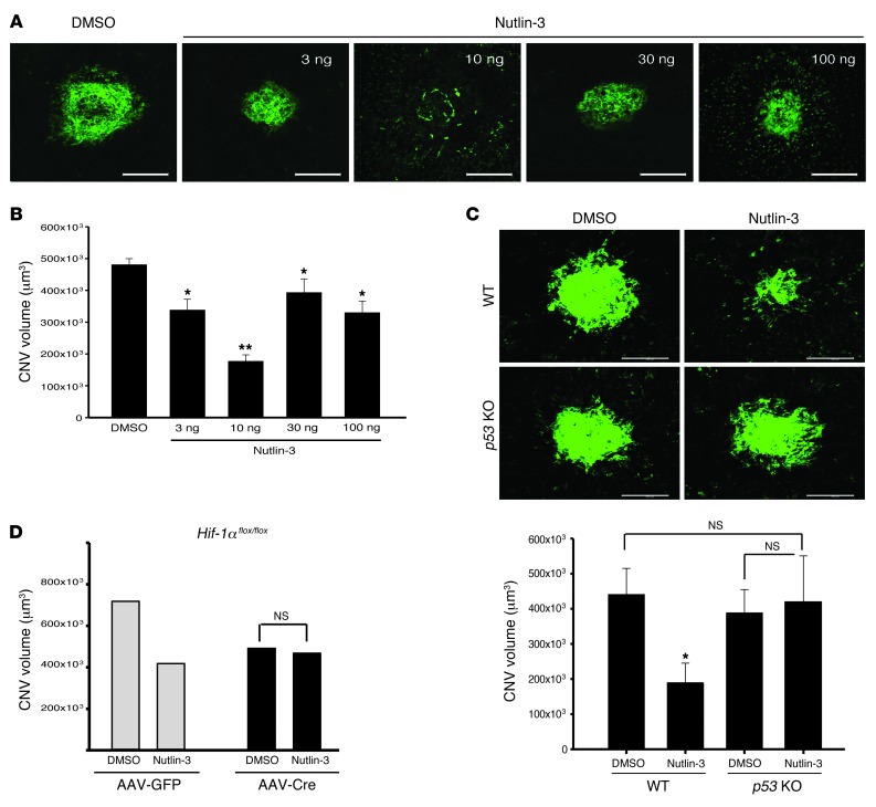
Figure 8. Nutlin-3 inhibits pathologic retinal angiogenesis through the p53 pathway.
(A) Stacked confocal image of representative laser-induced CNV lesions in racemic Nutlin-3– and DMSO-treated eyes. Racemic Nutlin-3 showed maximal activity at 10 ng/17.2 μM. (B) Quantification of z-stack confocal images of laser induced CNV in the respective conditions (n = 16–40). (C) Representative images of littermate controls (upper panels) after intravitreal injection of DMSO and racemic Nutlin-3. Similar experiments in p53–/– mice demonstrated no appreciable difference between DMSO- and Nutlin-3–injected mice. (n = 10–13). (D) Hif1aflox/flox mice were given either subretinal AAV1-VMD2-Cre or AAV1-VMD2-GFP. HIF-1α inhibition had no additional impact on Nutlin-3 antiangiogenesis (n = 6–10). Data represent mean ± SEM. *P < 0.05; **P < 0.0001; NS = P > 0.05. P = 0.052 for AAV1-GFP-DMSO vs. AAV1-GFP–Nutlin-3. Scale bars: 100 μm.

