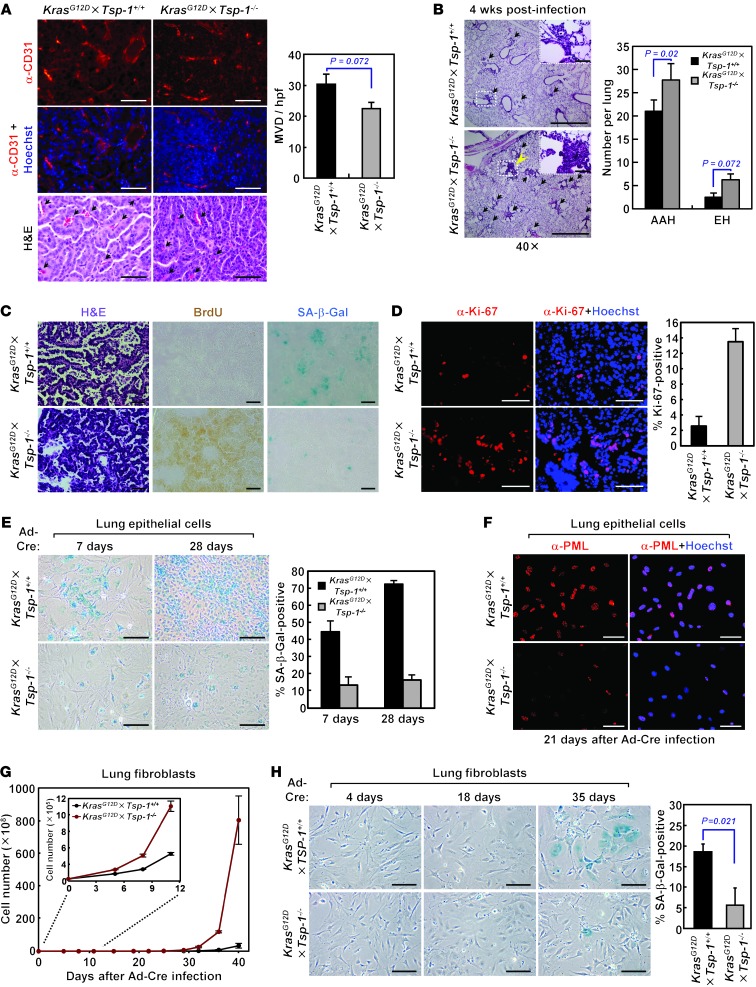
Figure 2. KrasG12D-induced senescence during lung tumor progression is compromised in the absence of TSP-1.
(A) Microvessel density in large lung adenomas of similar size from KrasG12D×Tsp-1+/+ and KrasG12D×Tsp-1–/– mice 21 weeks after oncogenic Kras activation. Tumor microvessels were visualized by immunostaining for CD31 and quantified by measuring microvessel density per high-powered field (MVD/hpf). Arrows indicate representative tumor microvessels. (B) Hyperplastic lesions in lungs 4 weeks after Ad-Cre inhalation. Lungs were isolated (n = 4 per group), and the number of lesions with atypical adenomatous hyperplasia or epithelial hyperplasia (EH) was counted after H&E staining. Arrows indicate hyperplastic lesions. (C) H&E staining, BrdU uptake, and SA–β-gal staining of serial sections from lung tumors harvested 21 weeks after Ad-Cre infection. (D) Proliferation in lung tumor lesions, as assessed by immunostaining for Ki-67, 21 weeks after Ad-Cre infection. (E) KrasG12D-induced senescence of primary adult lung epithelial cells isolated in vitro, as assessed by SA–β-gal positivity, at the indicated times after oncogenic Kras activation. (F) PML expression 21 days after oncogenic Kras activation in primary adult lung epithelial cells, determined by immunostaining. (G) Proliferation of primary adult lung fibroblasts isolated in vitro at the indicated times after oncogenic Kras activation. (H) KrasG12D-induced senescence of primary adult lung fibroblasts in vitro, measured by SA–β-gal positivity, at the indicated times after oncogenic Kras activation. SA–β-gal staining was quantified 35 days after Kras activation. Scale bars: 50 μm (A; B, insets; C; and D); 1 mm (B); 200 μm (E and H); 100 μm (F). Data represent mean ± SEM.

