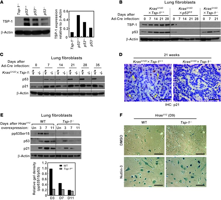
Figure 4. Oncogenic Kras activation triggers TSP-1 upregulation in a p53-dependent manner.
(A) TSP-1 protein expression in lungs harvested from p53+/+, p53+/–, and p53–/– mice. Isolated lung tissue was probed for TSP-1 expression by immunoblotting. Tsp-1–/– lung tissue served as negative control. TSP-1 expression relative to β-actin (loading control) was quantified by densitometry. (B) TSP-1 expression in the absence of p53 after KrasG12D activation in primary lung fibroblasts. KrasG12D×Tsp-1+/+, KrasG12D×p53fl/fl, and KrasG12D×Tsp-1–/– lung fibroblasts were isolated, infected with Ad-Cre to activate oncogenic Kras and delete p53, harvested at the indicated time points, and probed for TSP-1 expression by immunoblotting. (C) Western blot of p53 and p21cip1 expression in KrasG12D×Tsp-1+/+ and KrasG12D×Tsp-1–/– primary lung fibroblasts isolated at the indicated times after KrasG12D activation. β-actin served as loading control. (D) Immunohistochemistry for p21cip1 in KrasG12D×Tsp-1+/+ and KrasG12D×Tsp-1–/– lung sections isolated 21 weeks after KrasG12D activation. Arrows indicate p21cip1-positive cells. (E) pp53Ser15 after oncogenic Hras activation. Tsp-1+/+ and Tsp-1–/– lung fibroblasts were infected with HrasV12 retrovirus, harvested at the indicated days, and probed for pp53Ser15, p53, p21cip1, and β-actin by immunoblotting. Uninfected lung fibroblasts served as negative controls. pp53Ser15 expression relative to total p53 was quantified by densitometry. (F) Nutlin-3 treatment restored HrasV12-induced senescence in Tsp-1–/– lung fibroblasts. Lung fibroblasts were infected with HrasV12 or mock retrovirus, treated with 5 μM nutlin-3 or DMSO for 9 days, then stained for SA–β-gal to detect senescent cells. Scale bars: 50 μm (D); 200 μm (F).

