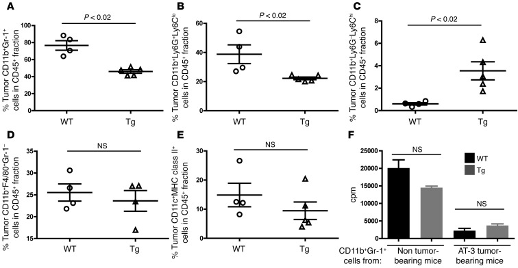
Figure 5. Intratumoral MDSC accumulation is also diminished in Irf8-Tg mice.
Flow analysis of the indicated myeloid populations from primary tumor tissue of WT or Irf8-Tg mice at endpoint, as shown in Figure 4, A or C. Total live cells were first gated on the CD45+ leukocyte fraction. The gated CD45+ fraction was then plotted in relation to the myeloid markers shown in a manner similar to that in Supplemental Figure 1. The data are illustrated for total MDSCs (A), MDSC subsets (B and C), macrophages (D), and total DCs (E). (F) Ability of CD11b+Gr-1+ cells from AT-3 tumor–bearing WT or AT-3 tumor–bearing Irf8-Tg mice to suppress T cell proliferation in response to immobilized anti-CD3 mAb. T cells (1 × 10⁵/well) and CD11b+Gr-1+ cells (5 × 10⁴/well) (n = 3 determinations; *P < 0.01; T cells + anti-CD3 mAb without any CD11b+Gr-1+ cells, 23,212 ± 1,865).

