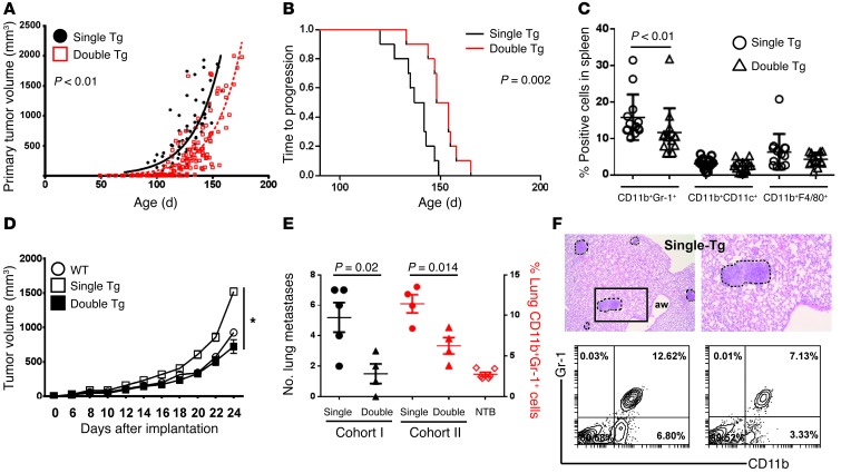
Figure 7. Irf8 enhancement slows autochthonous tumor growth.
(A) Tumor growth rate in double-Tg versus single-Tg mice, as determined after tracking the single largest tumor. Each symbol denotes a tumor measurement in a single mouse over time (n = 13 mice each; all Irf8-Tg mice from line 370). (B) Kaplan-Meier plot of the data in A, based on time to progression to approximately 50% of maximal tumor growth as a surrogate endpoint. (C) Each data point represents flow analysis of the indicated myeloid subset from single-Tg or double-Tg mice at endpoint. (D) Myeloid/tumor admixture experiments, as in Figure 2. AT-3 cells were mixed with splenic CD11b+Gr-1+ cells recovered from single-Tg, double-Tg, or non-tumor-bearing mice (WT), and tumor growth was recorded. The cells from single-Tg mice, but not those of double-Tg mice, showed a significant (*P < 0.001) increase in the AT-3 tumor growth rate relative to cells from WT mice (n = 5 mice/group; one of 2 separate experiments). The data are expressed as the mean ± SEM for the indicated number of mice. (E) Quantification of lung metastasis using histology (termed cohort 1) or CD11b+Gr-1+ cell frequency using flow cytometry analysis of total lung digests (termed cohort 2) from single-Tg or double-Tg mice. NTB, non-tumor-bearing WT mice. Both cohorts were matched based on age and primary tumor burden, as indicated in Results. (F) Representative H&E-stained images of metastatic foci in MTAG mice (upper left, original magnification, ×40; upper right, original magnification, ×100) and flow plots of CD11b+Gr-1+ cell populations (bottom panel).

