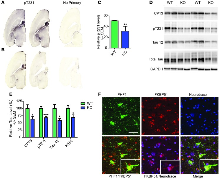
Figure 1. Tau expression is decreased in the Fkbp5–/– mice and associates with FKBP51 in the AD brain.
Representative images of brain tissue from (A) wild-type and (B) Fkbp5–/– mice stained for pT231 tau and corresponding sections without primary (no primary). Scale bar: 2,000 μm. (C) Quantification of pT231 stain (± SEM). **P = 0.0097. (D) Representative Western blot of brain homogenates from Fkbp5–/– mice probed for CP13 (pS202/pT205), Tau 12 (total tau), pT231 tau, H150 (total tau), and GAPDH. (E) Quantification of duplicate Western blots (± SEM). *P < 0.05 for CP13, Tau 12 (total tau), and H150 (total tau); ***P < 0.001 for pT231. (F) Fluorescent micrograph of human cortex stained with FKBP51, PHF1 (pS396/pS404), and Neurotrace (neuronal nuclei) antibodies (original magnification, ×60). Scale bar: 50 μm.

