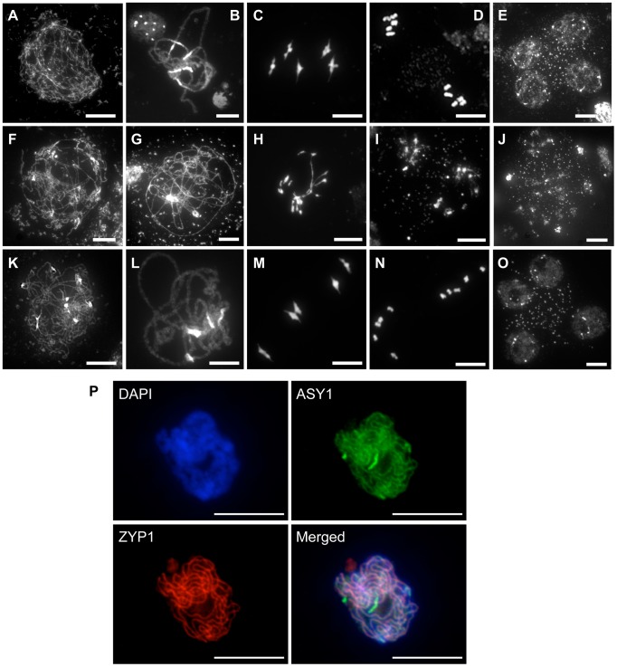
Figure 2. Progression of meiosis in WT (A to E), rad51/rad51 (F to J) and rad51/rad51 plants expressing the RAD51-GFP fusion protein (K to O).
DAPI staining of pollen mother cells (PMC) during meiosis. (A, F, K), Leptotene, (B, G, L) Pachytene, (C, H, M) Metaphase I, (D, I, N) Metaphase II and tetrad showing four meiotic products (E,O) and the equivalent stage with heterogeneous DNA masses in the rad51 mutant (J). (Scale Bar: 10 µm). (P) Immunolocalisation in rad51/rad51 RAD51-GFP PMC shows that synaptonemal complex (SC) proteins ASY1 (SC axial element) and ZYP1 (SC transverse filament) are correctly loaded along chromosome axes at pachytene indicating normal completion of synapsis. DAPI (blue), ASY1 (green), ZYP1 (red) and merged images are shown. (Scale Bar: 10 µm).

