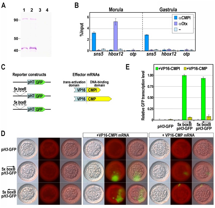
Figure 3. Association of the CMPl protein to the boxB cis-element on sns5.
(A) Western blot analysis to test the specificity of the anti-CMPl antibody. Nuclear extracts from morula (lanes 1, 3) and gastrula (lanes 2, 4) embryos were fractioned by SDS-PAGE, blotted on nitrocellulose membrane and incubated with anti-CMPl antibody (lanes 1, 2) or pre-immune serum (lanes 3, 4). (B) ChIP-qPCR analysis of the sns5 occupancy by CMPl. ChIP assays were performed on chromatin extracted from embryos at the indicated stages and precipitated with antiserum against CMPl or Otx, or incubated without adding antibodies (−), followed by qPCR amplification of an sns5 fragment containing the boxB sequence, or hbox12 and otp promoter fragments. As described previously, the association of the Otx transcription factor to its binding site within the hbox12 promoter correlates with the hbox12 transcriptional state [32]. Data are normalized according to the percent of input method. Bars are as in Figure 2D. (C) Scheme of the reporter and effector constructs used in the trans-activation assay. (D) Trans-activation analysis in developing sea urchin embryos observed at the mesenchyme blastula stage. DIC, epifluorescence and merged images, respectively ordered from top to bottom, are shown for each embryo. (E) GFP expression levels assessed by qPCR in transgenic embryos at the mesenchyme blastula stage. Graphs show n-fold changes in mRNA expression level of GFP based on the normalized threshold cycle numbers, with respect to the 5×boxB-pH3-GFP/VP16-CMPl co-injected embryos. Bars are as in Figure 2D.

