
Figure 5. Known conformations of reactive peptides.
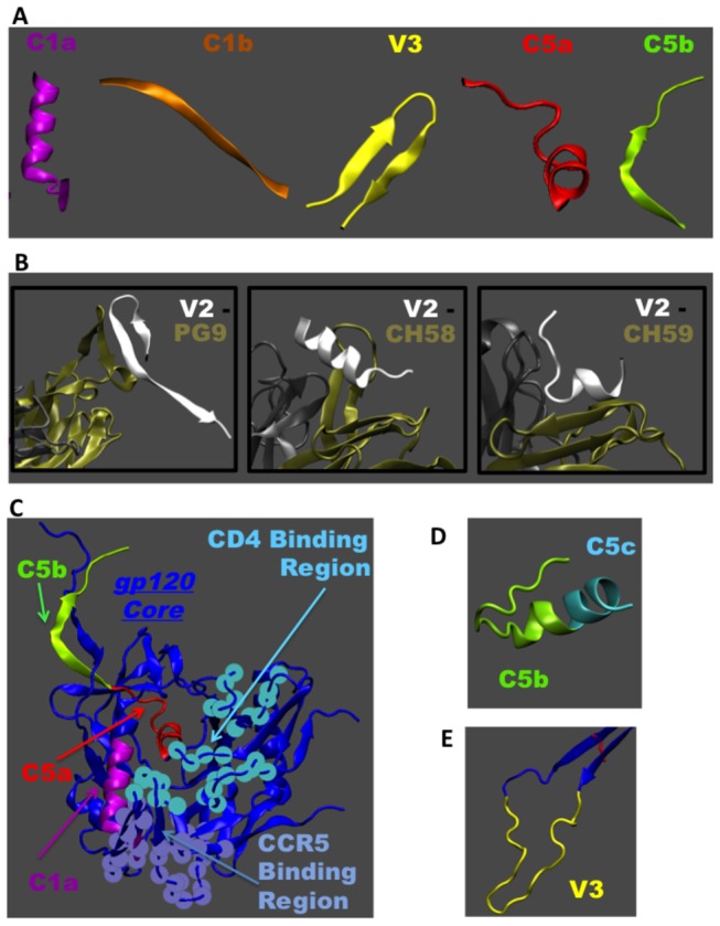
A. Conformations of reactive peptides known from solved X-ray structures of gp120 core or fragments of gp120 in complex with antibodies. B. V2 peptide is shown with respect to different conformations it adopts depending on the bound antibody. Structures with antibodies PG9 (PDB: 3U4E), CH 58 (PDB: 4HPO) and CH 59 (PDB: 4HPY) are shown where tan and gray colors indicate the light and heavy chains of the antibody, respectively. C. The CD4 bound structure of gp120 with N-and C- terminal regions is used as a template to show the C1a, C5a, C5b peptides in the context of entire gp120 monomer structure (PDB: 3JWD). Also shown are the CD4 and CCR5 binding regions respect to these peptides. D. NMR structure of isolated region of C-terminal end of gp120 showing C5b and part of C5c peptides (PDB: 1MEQ). E. V3 peptide is shown with respect to the rest of the gp120 core (PDB: 2B4C).