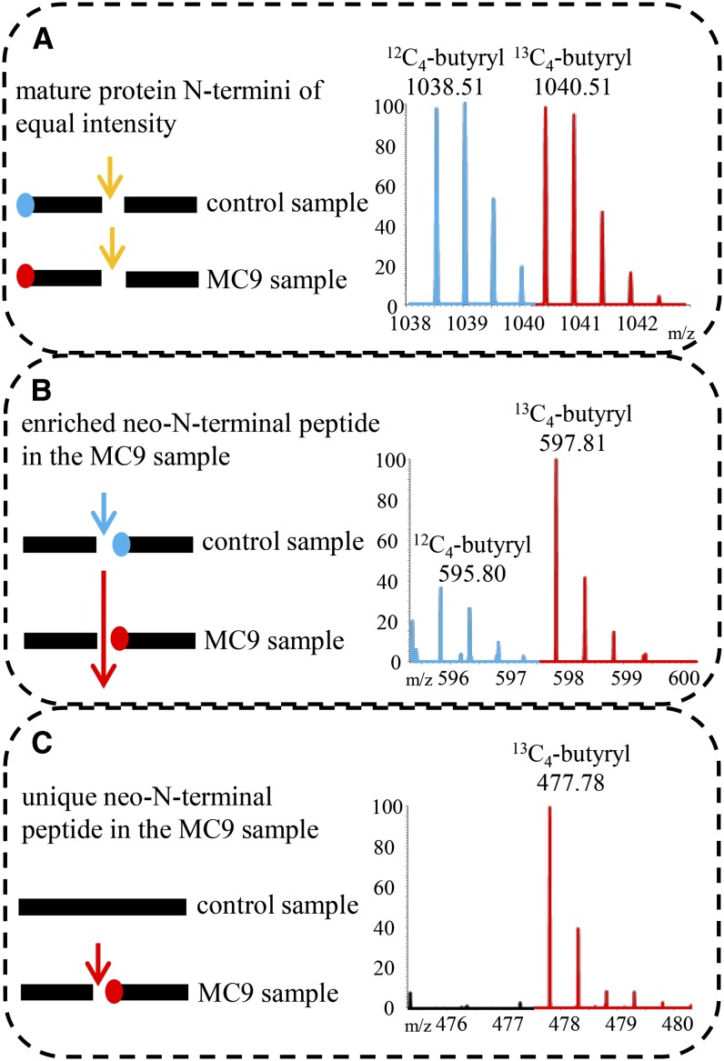
Figure 2.
Illustration of the Different Categories of Isolated (Neo)-N-Terminal Peptides.
Mass tagging allowed the differential comparison of N-terminomes derived from samples in which MC9 was active (Col-0 or 35S:MC9 or added rMC9) or absent (control sample mc9). MS spectra of N-terminal peptides were, in most cases, identified as doublet ions of light (in blue) and heavy (in red) isotopes. m/z, mass-to-charge ratio.
(A) Protein cleaved in both samples to the same extent (i.e., by trypsin; yellow arrow) and fragments bearing the mature protein N terminus. As the amounts of these peptides were approximately equal, no MC9-driven proteolysis could be deduced.
(B) Protein cleaved in both samples, but at a higher frequency in the MC9 sample (large red arrow). The neo-N-terminus fragment was generated in both samples, but in significantly higher amounts in the MC9 sample. Proteolysis of the target in the control sample might have occurred by a redundant protease activity (blue arrow). The fragment is identified as a doublet with the highest intensity ion derived from the MC9 sample.
(C) Protein solely cleaved by MC9 (red arrow) in the samples of active MC9 proteases. Thereby, a fragment with a neo-N-terminus was generated, later identified as unique in the MC9 N-terminome and, thus, as a singleton ion.

