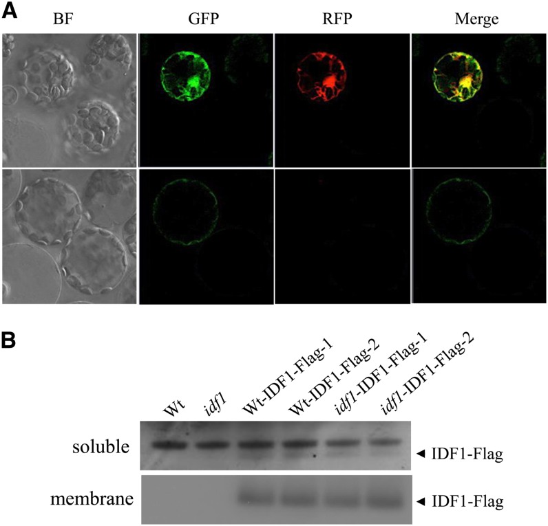
Figure 6.
Subcellular Localization of IDF1.
(A) Subcellular localization of IDF1 in protoplasts. 35S:IDF1-GFP and 35S:mCherry were cotransfected into Arabidopsis protoplasts, and the transient expression of the fluorescent reporter proteins was detected by confocal microscopy. Two sets of images of different focal planes (top and bottom rows) represent the bright-field (BF) GFP-specific signal for IDF1-GFP (excitation, 488 nm; emission, 500 to 530 nm), the red fluorescent protein (RFP)–specific signal for mCherry (excitation, 561 nm; emission, 575-630 nm), and a merged image of the GFP and red fluorescent protein images.
(B) IDF1-Flag was detected with anti-Flag antibody in both the soluble and membrane fractions of root proteins from the transgenic wild-type (Wt) and idf1-1 (idf1) expressing IDF1 promoter:IDF1-Flag (Wt-IDF1-Flag-1,-2 and idf1-IDF1-Flag-1,-2). Two representatives of at least three lines of each transgenic construct characterized are shown.

