Figure 7.
IDF1 Directly Interacts with IRT1.
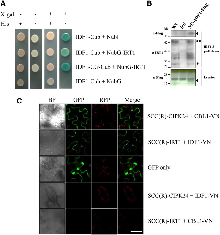
(A) The interaction between IDF1 and IRT1 was detected using in the DUALmembrane yeast two-hybrid system. IDF1 was used as the fused bait protein (IDF1-Cub and IDF1-CG-Cub). IRT1 was used as the fused prey protein (NubG-IRT1). The presence (+) or absence (−) of His or 5-bromo-4-chloro-3-indolyl-β-d-galactopyranoside (X-gal) are indicated.
(B) The antibody IRT-U was used to pull down the protein complex with IRT1 from the wild type (Wt), irt1, and IDF1-Flag overexpressor (35S-IDF1-Flag) extracts. Anti-Flag (M2, αFlag) and anti-IRT1 (IRT1-U, αIRT1) antibodies were used to detect the target proteins in the IRT1-U pull-down sample and lysates. Arrowheads indicate IDF1-Flag, the single dot indicates IRT1, and the double dot indicates IRT1-Ub4.
(C) The interaction between IDF1 and IRT1 [SCC(R)-IRT1/IDF1-VN] was observed by BiFC conducted in the leaves of N. benthamiana. The green fluorescence of SCC(R)-CIPK24/CBL1-VN is presented as a control for interaction at the plasma membrane, and GFP shows the cytoplasm. The combination of SCC(R)-CIPK24/IDF1-VN and SCC(R)-IRT1/CBL1-VN were used for false-positive tests. BF, bright field. Bar = 50 μm.

