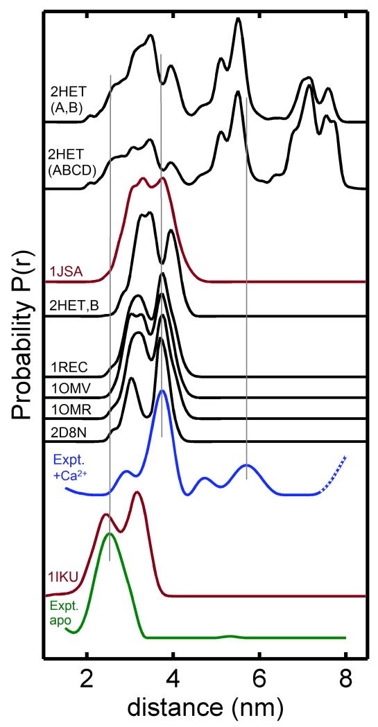
Figure 3.
Distance distributions for spin-labels attached at C39 and N120C were calculated using NMR structures of recoverin (1IKU and 1JSA, highlighted in red) and several x-Ray crystal structures (1REC, 1OMV, 1OMB, 2HET; in black). Experimental distance distructions calculated from the DEER data are shown for Ca2+-free recoverin (green) and Ca2+-bound recoverin (blue).

