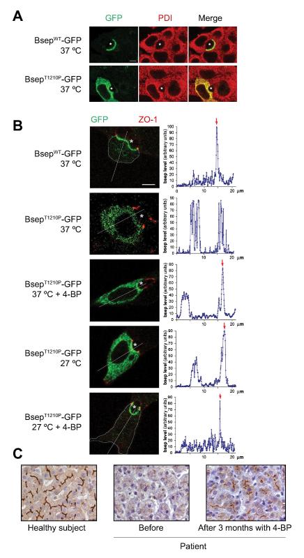
Figure 3.
In vitro studies of mutant BsepT1210P–GFP in a polarize cell line and the effect of 4-PB in a PFIC2 child expressing this mutation. (A) Immunofluorescent localization of GFP constructs of WT and mutant BSEP (green) and protein disulfide isomerase (PDI) (red) in Can 10 cells. (B) 4-PB and low temperature increase canalicular membrane expression of BsepT1210P–GFP (green). The apical membrane was delineated with immunofluorescent labeling of the tight junction protein, zonula occludens 1 (ZO-1) (red). Quantification of Bsep–GFP levels is shown in graphs to the right. Red arrows indicate canalicular fluorescence reinforcement, when present. A thin blue-grey line was drawn on confocal images to delineate cell limits, when they were hardly visible. * canaliculus; bar: 5 μm. (C) BSEP immunostaining of human liver sections: healthy subject (left), patient before (middle) and after 3 months with 4-PB therapy (right). Bar: 30 μm. (Reproduced with permission from (Gonzales et al., 2012a)).

