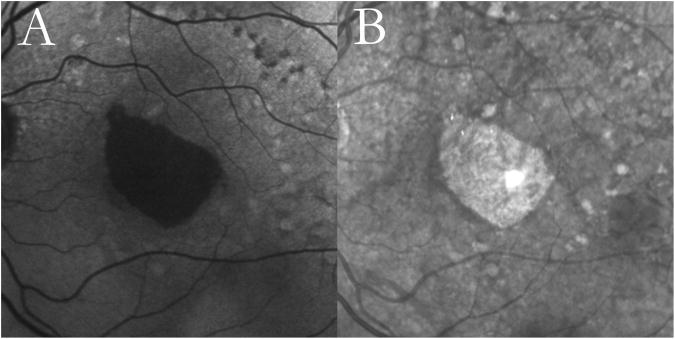
Figure 1. Unilobular geographic atrophy (GA) with reticular macular disease (RMD).
Autofluorescence (A) and infrared (B)images from a 68-year-old female with age-related macular degeneration showing unilobular GA. Both modalities show RMD around the atrophic lesion and temporally to the optic disc, as well as large soft drusen more peripherally.

