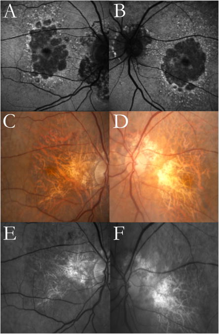
Figure 8. Multilobular geographic atrophy (GA) demonstrated in autofluorescence (AF) imaging but not in color fundus (CF)or red free (RF) photography.
Images from an 88-year-old female with age-related macular degeneration. Multilobular GA and reticular macular disease (RMD) around the atrophic lobules are clearly visible in the AF images (top row). The CF (middle row) and RF (bottom row) images do not show the multilobular nature of the GA but do show RMD superotemporally in both eyes. Peripapillary atrophy is present in both eyes in all image modalities.

